Figures & data
FIG. 1 Schematic diagram of the experimental equipment used to produce Si nanopowders.
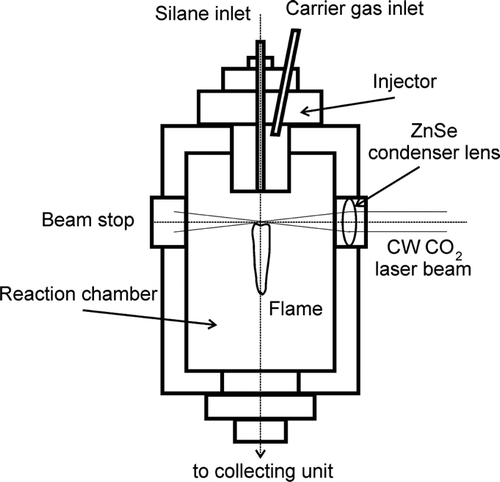
FIG. 2 TEM micrographs of the Si nanopowders Si 1 (a), Si 2 (b), and Si 3 (c) produced at 840°C, 1030°C, and 1400°C, respectively.
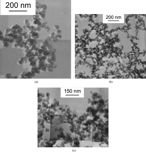
TABLE 1 Experimental data from TEM and XRD characterization of the silicon nanopowders produced by laser pyrolysis
FIG. 3 X-ray diffraction pattern recorded on the sample Si 3 with Cu-kα (= 0.154 nm) incident radiation. There is no evidence of amorphous Si phase formation.

FIG. 4 Normalized line profiles of the Si (111) reflection peaks for samples Si 1 and Si 3. The full width at half maximum of the peaks is inversely proportional to the crystal coherence length or crystallite size.
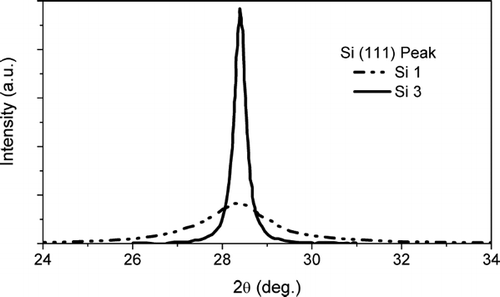
TABLE 2 Average particle diameter calculated by the classical analytical model after a reaction time of 9 ms and ratio between the characteristic coagulation time to the sintering time
TABLE 3 Summary of the selection rules for particle collisions in modelling the PSD and CSD evolution during coagulation
FIG. 5 Evolution of the PSD and CSD average diameters calculated by the modified sectional model for a reaction time of 9 ms.
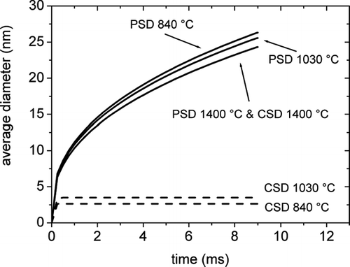
FIG. 6 Calculated behaviour of the average particle size of PSD and CSD with temperature and comparison with the experimental data from TEM and XRD. The dashed line represents the limiting size for the liquid to solid transition.
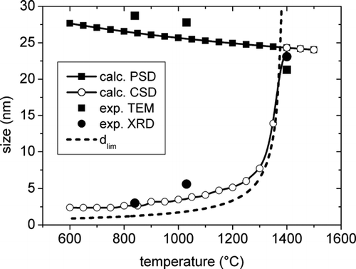
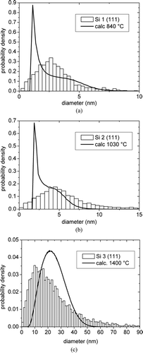
FIG. 7 Comparison between the calculated PSDs and the distributions from TEM measurements for the samples (a) Si 1, (b) Si 2, and (c) Si 3.
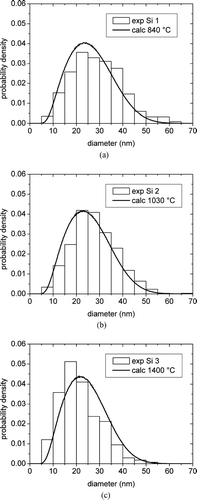
FIG. 8 Comparison between the calculated CSDs and the distributions from XRD measurements on Si (111) peaks for the samples (a) Si 1, (b) Si 2, and (c) Si 3.