Figures & data
FIG. 1 Schematic of the Aerosol Mass Spectrometers. The lower part shows the vacuum system that is identical in both instruments, the upper parts show the mass analyzers and the detection system of Q-AMS (left) and ToF-AMS (right).
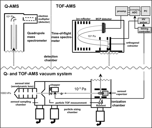
FIG. 2 Flow chart of the ToF-AMS data analysis procedure. Analysis of MS mode data is covered in the left column, of PToF mode data in the right column. Shown in the middle gray column are the corrections and calculations which are applied to both types of data.
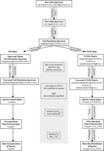
FIG. 3 Visualization of the peak integration algorithm. All points between the solid lines (−0.3 to +0.2 m/z around the exact m/z of a peak) belong to the peak integration area. In order to account for baseline drifts over the peak, the baseline of each peak is defined as the average value of the baseline range before (Baseline Area 1) and after (Baseline Area 2) the peak. The resulting area of the peak is calculated as the sum of all values within the integration area minus its average baseline value.
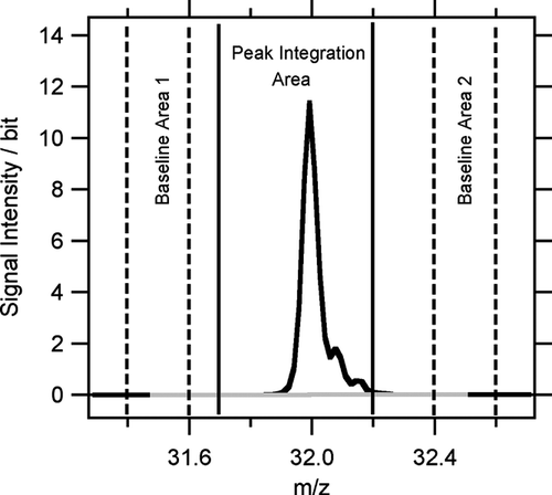
FIG. 4 Time series of non-refractory nitrate, sulfate, ammonium, total organics and total non-refractory mass concentrations, measured with ToF-AMS (upper panel) and Q-AMS (lower panel).
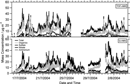
FIG. 5 Correlations of ToF-AMS and Q-AMS mass concentrations for (a) nitrate, (b) sulfate, (c) ammonium, and (d) organics. Grey markers indicate data from period I (July 16–July 25 17:00 h), black markers from period II (July 27 12:00 h–August 4 9:00 h). The solid lines are linear fits for period I and II, the dashed line is the 1:1 line.
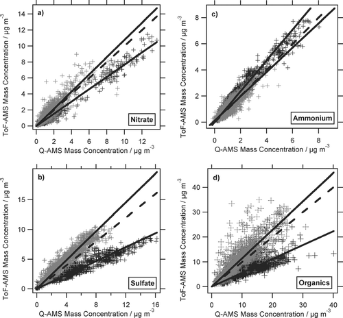
TABLE 1 Parameters of the linear fits in
TABLE 2 Parameters of the average species-resolved size distributions, obtained by log-normal fits ( and ). Mode diameter values are given in nm; distribution widths are given as GSD
FIG. 7 Modeling input and output of threshold-related species concentration changes. Top panel: distributions of single and small multiple ion events as a function of ion signal intensity; middle panel: loss of single and small multiple ion events as a function of threshold position; lower panel: resulting modeled species concentration change as a function of threshold position. Signal intensity and threshold position are normalized to the average single ion signal intensity.
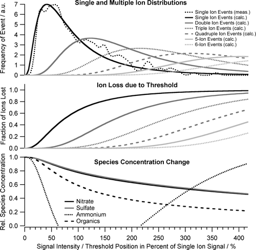
FIG. 6 Average mass spectrum in ions per second (lower graph) and average modeled fraction of signal arriving at the detector as single ions per mass spectrum (top graph). The larger the single ion signal fraction is the more sensitive the individual ions are to threshold-related small ion signal loss.
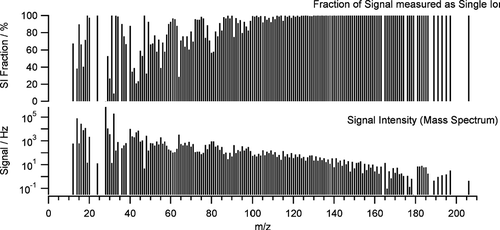
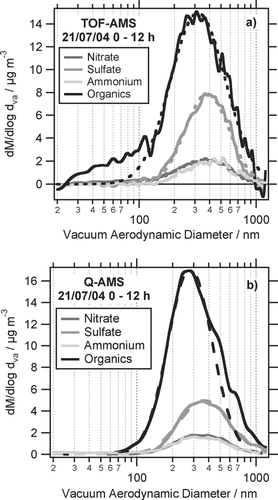
FIG. 8 Average size distributions for July 20 of nitrate, sulfate, ammonium, and organics, measured with (a) ToF-AMS and (b) Q-AMS together with log-normal fits to the distributions.
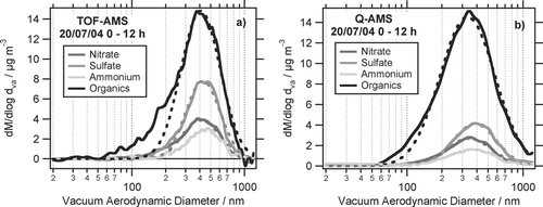
FIG. 9 Same as but for July 21.