Figures & data
FIG. 1 The ambient aerosol size distribution overlaid with three biological aerosol populations.
FIG. 2 Schematic and photo of the cyclone with its water vortex.
FIG. 3 Flow configuration to atomize monodisperse PSL particles and measure the total number concentration to calibrate the cyclone collection efficiency.
FIG. 4 Airborne monodisperse PSL particles as scanned by the SMPS and APS. The 50 nm size began to combine with the size distribution of the atomized water.
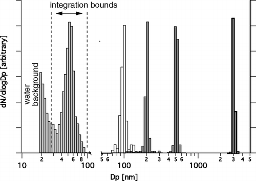
FIG. 5 Measured emission intensities by the fluorimeter for a series of dilutions from each monodisperse PSL particle solution.
TABLE 1 Calculated uncertainty in predicted concentration based on the variance in the linear regression
TABLE 2 Characteristics of the virus particles used in this study
FIG. 6 The flow configuration to stain and detect the BLV inline with the Partec CyFlow® space flow cytometer.
FIG. 7 The flow configuration to generate and collect an airborne virus aerosol. The online staining and cytometric detection was performed with the Partec CyFlow® space. Offline collection of samples and cytometric detection was performed with the Becton-Dickinson FACS Calibur.
FIG. 8 The collection efficiency curves for the cyclone in stand-alone mode and preceded by aerosol growth by water vapor condensation.
FIG. 9 Stained media and stained Baculo-virus stock solutions analyzed on (a) a Becton-Dickinson FACS Calibur and (b) a Partec CyFlow® space.
FIG. 10 Increasing emission of (a) the stained Baculo-virus population, and (b) the peak emission intensity of the virus population versus time.
FIG. 11 Offline versus inline staining and detection of the Baculo-virus. Virus for inline staining was diluted by a factor of 10.
FIG. 12 (a) The atomized BLV collected by the cyclone (“sample”) and compared against total filtered air (“background”) and the inline staining result. (b) The “sample” collected by the cyclone replotted against the forward scatter parameter to separate the fluorescence background of the bubbles.
FIG. 13 Airborne TMV collected by the cyclone and compared against background room air and TMV stock. All samples were stained with (a) SYTO-9 and (b) SYBR-Gold.
FIG. 14 ESEM images of (a) a freeze-dried aliquot of the virus stock solution, and (b, c) an aliquot of two independent samples of airborne TMV collected by the cyclone with aerosol growth.