Figures & data
FIG. 1 Schematics of SP-AMS instrument that show the particle beam, laser beam, and ion extraction axes and define the horizontal and vertical axes of the instrument as discussed in the text. (a) Top-down view. (b) Cross-section view looking up the particle beam axis.
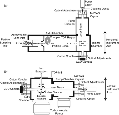
FIG. 2 Cartoon of coated soot particles passing through laser beam (from left to right), with transit times of 5–20 μs (dependent upon size). As the coated soot particle heats, the top plot shows measured light scattering (red) and incandescence signals (blue) as observed by an SP2 (Gao et al. Citation2007), and the bottom schematic shows the vaporization of nonrefractory species (R'(g)) and refractory carbon clusters (C m(g)) and the resulting ions formed from electron impact ionization in the detection region of the SP-AMS. Subscripts s and g denote solid and gas phase, respectively.
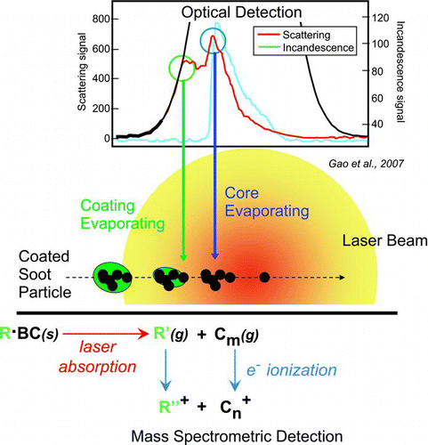
FIG. 3 Mass spectrum of denuded (250°C) ethylene flame soot averaged over 20 min. Carbon ion clusters are shown in black and organic signals (i.e., C x H y and C x H y O z ) are shown in green. Significant carbon ion clusters are spaced 12 m/z apart (i.e., C1) for m/z < 384 and 24 m/z apart (i.e., C2) for m/z > 384. The first inset highlights the stable carbon ion cluster sequence for m/z greater than 384 (i.e., C32 +), which are fullerene structures. The second inset shows the measured 13C isotopic distribution (black; raw signal) compared with NIST values (red sticks) for the stable C60 cluster, buckminsterfullerene. The pie chart shows the relative abundance of the carbon ion clusters for C1–C5 (49%), C6–C31 (28%), and C32–C85 (24%).
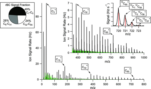
TABLE 1 SP-AMS measured particulate species for the 3 vaporizer combinations
FIG. 4 Laboratory generated nascent soot sampled by a SP-AMS with both laser and tungsten vaporizers. The mass spectra in (a) show the laser-on, laser-off, and the difference (laser-on – laser-off) conditions. Carbon ions are shown in black, organic ions in green, and CO2 + ion signal is highlighted in violet in the difference spectrum. The integrated ion signals for refractory carbon, organic, and CO2 + (including fragmentation table entries derived from CO2 + ion signals) are shown in (b) as a function of experiment time.
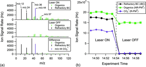
FIG. 5 Size distributions for uncoated (a) and DOS oil coated (b and c) monodisperse 225 nm particles generated by atomizing and size selecting 50 nm glassy carbon spheres from a water suspension. The carbon ion signals (black) are plotted on the left axes and the organic ion signals (green) are plotted on the right axes, both normalized to the log10 size bin width (i.e., dS/dlogd va). The vertical line through the mode of the uncoated particle distribution is to emphasize shift in d va with added DOS. The coating thickness estimates come from measured mobility diameters.
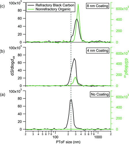
FIG. 6 SP-AMS rBC calibration. (a) SP-AMS integrated carbon ion signals per particle versus the per particle mass as measured by a CPMA for atomized Regal black and nascent and denuded (250°C) ethylene flame soot particles. The change in slope (Regal black < nascent < denuded) is related to collection efficiency differences caused by greater particle beam divergence for nonspherical particles. See text for details. (b) Normalized sensitivity curve for the SP-AMS (carbon ion signal per rBC particle mass) for uncoated (red) and DOS coated (black) 250 nm Regal black particles as a function of the laser power measured passing through the output coupler mirror. The normalized DOS organic ion signal per particle mass for the coated particles (green) saturates at lower laser power than the refractory carbon ion signals as expected due to the less refractory nature of DOS.
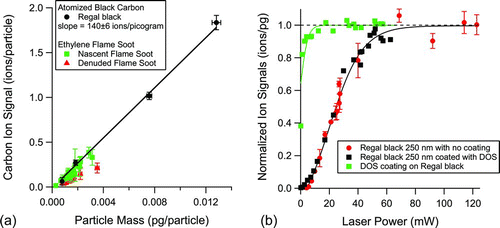
FIG. 7 Average mass spectrum (Hz) for ambient data sampled using a SP-AMS (laser vaporizer only) from a busy urban street in Chestnut Hill, MA. The figure shows the relative ion signals for rBC (black) and the nonrefractory chemical species, organics (green), sulfates (red), nitrates (blue), and ammonium (orange), on the black carbon particles. The first inset shows the measured fullerene ion series observed in ambient data. The pie chart shows the relative abundance of the carbon ion clusters for C1–C5 (86%), C6–C31 (10%), and C32–C70 (4%).
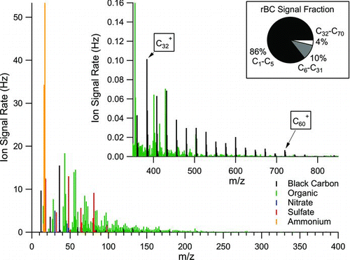
FIG. 8 Time dependent ambient data obtained with the SP-AMS (laser vaporizer only) and a MAAP black carbon absorption instrument in Chestnut Hill, MA. The SP-AMS and MAAP black carbon measurements (a) are correlated (R 2 = 0.76) and show multiple aerosol plumes containing black carbon particles. The SP-AMS nonrefractory organic (b) and inorganic (c) particulate mass associated with rBC particles are shown to be dominated by organic species. Two broad plumes (∼1.5 h) are highlighted; one plume is dominated by black carbon mass and the other by nonrefractory organic mass.
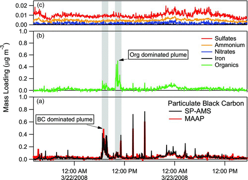
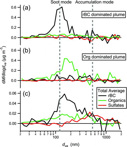
FIG. 9 SP-AMS average size distributions (mass loading versus vacuum aerodynamic diameter) of rBC, organics (Org), and sulfates (SO4) for (a) “rBC dominated” plume (highlighted in Figure 8), (b) “Org dominated” plume, and (c) as an average for the total sampling period.