Figures & data
FIG. 1 Deposition chamber schematic. Dry powder pmma particles were dispersed, electrostatically neutralized, and deposited on test wafers. Aerosol concentration and aerodynamic particle size were measured directly with an APS. Average electrostatic charge per particle was measured with a Faraday cage filter sampler and electrometer.
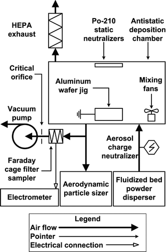
FIG. 2 Particle resuspension test schematic. Particle contaminated substrates were vibrated with an ultrasonic transducer within a humidity controlled chamber. Particle resuspension was recorded with a high speed camera through a bright field microscope. Surface vibrations were measured directly with LDV.
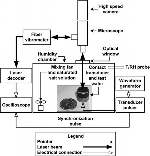
TABLE 1 Saturated salt solutions, and respective theoretical relative humidity conditions at saturation, used to vary capillary condensation during experiments. Saturation conditions were taken from Rockland (Citation1960)
FIG. 3 Surface roughness height (nm) vs. stylus profilometer linear scan distance, x (μm), for (a) TiO2, and (b) SiO2 wafer surfaces where 10 and 1 μm particles are drawn for TiO2 and SiO2 scans, respectively. Peak-to-peak distance is one the order of 1 μm, thus, particles effectively interact with a molecularly smooth surface rather than multiple surface asperities.
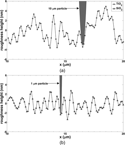
FIG. 4 Normalized particle size distributions measured with the APS and converted to physical diameter, dp .
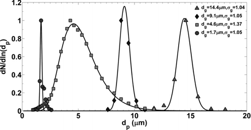
FIG. 5 Digital microscope image of test substrate contaminated with 14.4 μm pmma microspheres. Surface area coverage is approximately 10% where only monomers were included in the analysis of microparticle resuspension.
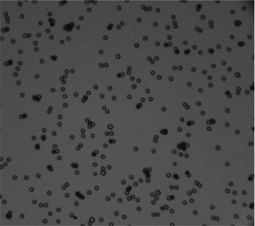
FIG. 6 Average charge per particle, n (electrons), calculated from APS measurements of deposition chamber particle concentration and electrometer measurements of Faraday cage filter current. Error bars represent standard deviations from current measurements. Data were fit with a dp 2 power law function.
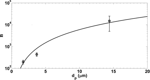
FIG. 7 (a) Wafer surface displacement (nm) measured with LDV and curve fit, (b) calculated surface velocity (m/s), and (c) calculated surface acceleration (m/s2) for 5 MHz contact transducer pulsed at maximum power (300 V).
FIG. 8 Percent microparticle resuspension from TiO2 surface as a function of the resuspension force normalized by the theoretical capillary force, EquationEquation (5), for data above 60% relative humidity. A sigmoid function was used to fit the linearized data set and find the mean response and 95% prediction interval bounds. Vertical error bars (±5%) are due to image processing and particle counting. Horizontal error bars were calculated with the Kline–McClintock equation from uncertainties in parameters used to calculate the capillary force.
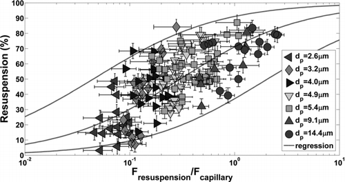
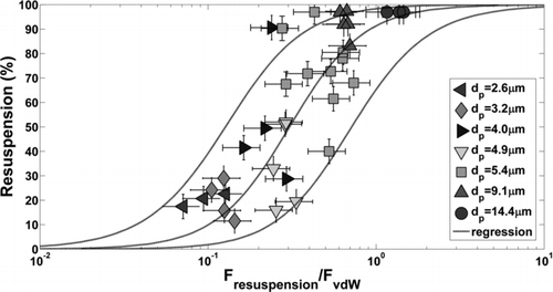
FIG. 9 Percent microparticle resuspension from TiO2 surface as a function of the resuspension force normalized by vdW force (JKR), EquationEquation (8), for experimental data below 60% relative humidity. The theoretical work of adhesion was optimized so that the midpoints of the regression curves (50% resuspension) for F*vdw and F*cap were equivalent. Vertical error bars (±5%) are due to image processing and particle counting. Horizontal error bars (±30%) were specified according to estimated uncertainty in the experimental work of adhesion.