Figures & data
FIG. 1 The computational domain of the study. (a) A multiblock structured grid with the adoption of the “butterfly” topology representing G3–G4 bifurcation of the human lung, (b) an illustration of the internal grid.
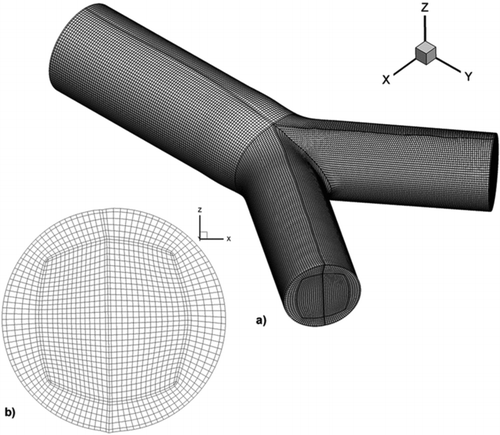
TABLE 1 Relative square error comparison for three consecutively finer computational domains
FIG. 2 Contour of the dominant velocity magnitude at the plane symmetry of PBR for the HFOV case for four different time phases (a) inhalation start, (b) inhalation peak, (c) flow reversal, and (d) exhalation peak.
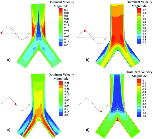
TABLE 2 Summary of flow characteristics for HFOV and NB cases
TABLE 3 Spherical particle diameters and Stokes number
FIG. 3 Contour of the dominant velocity magnitude at the plane symmetry of PBR for the NB case for four different time phases (a) inhalation start, (b) inhalation peak, (c) flow reversal, and (d) exhalation peak.
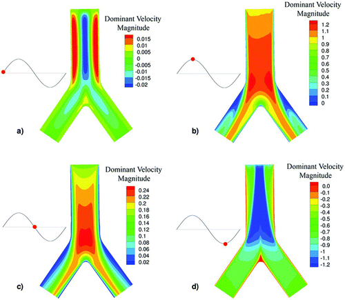
FIG. 4 Surface streamlines and secondary velocity contour magnitudes at a plane near the exit of the G3–G4 bifurcation at the flow reversion phase for (a) HFOV and (b) NB cases.

FIG. 5 Contour of particle deposition for 900 nm particles, around the area of the carinal ridge for the HFOV case at different time phases.
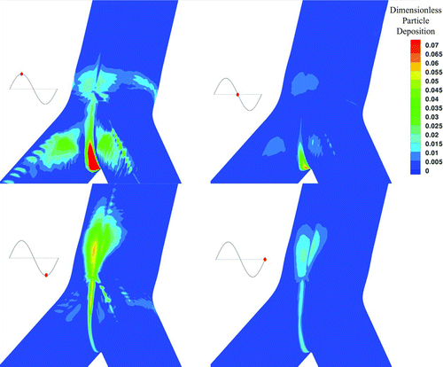
FIG. 6 Contour of particle deposition for 900 nm particles, around the area of the carinal ridge for the NB case at different time phases.
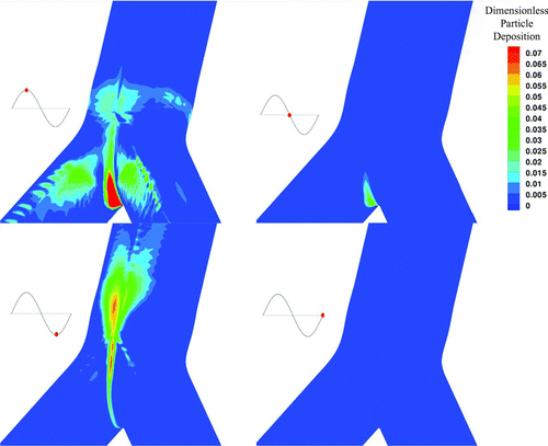
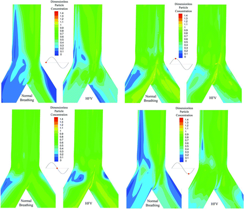
FIG. 7 Contour of particle concentration (particle diameter equal to 900 nm) at the cell centre adjacent to the wall for the HFOV and the NB case at different time phases.