Figures & data
FIG. 1. Comparison of fungal and bacterial concentrations using plastic and glass dishes. The top and bottom end of the box represent the 75th and 25th percentiles, respectively, and the line inside the box indicates the median. The bottom and top lines indicate 5th and 95th percentiles. Single points indicate the extremum values.
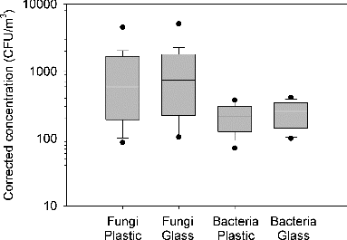
FIG. 2. Count median diameters (CMDs) of airborne fungi and bacteria with plastic and glass dishes. The top and bottom end of the box represent the 75th and 25th percentiles, respectively, and the line inside the box indicates the median. The bottom and top lines indicate 5th and 95th percentiles. Single points indicate the extremum values.
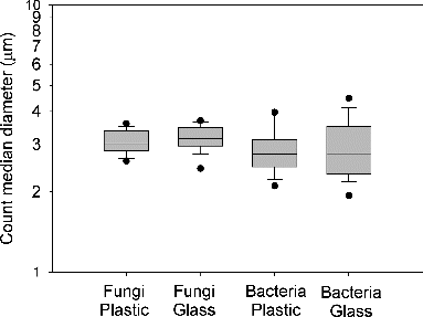
FIG. 3. Correlation of fungal concentrations using plastic and glass dishes.
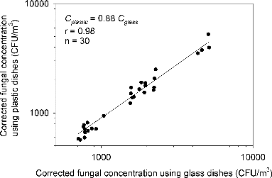
FIG. 4. Correlation of bacterial concentrations using plastic and glass dishes.
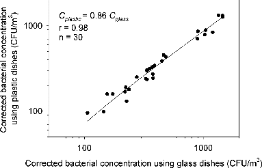
FIG. 5. Correlation of fungal concentrations obtained from colocated samples with plastic dishes (upper plot) and glass dishes (lower plot).
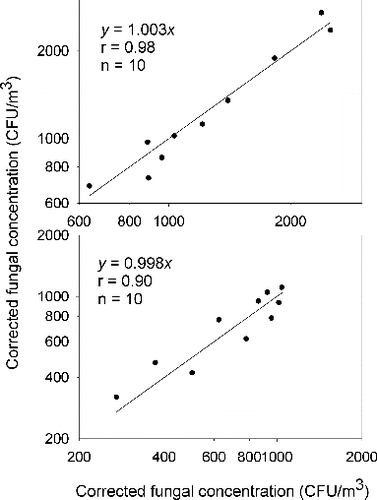
FIG. 6. Correlation of bacterial concentrations obtained from colocated samples with plastic dishes (upper plot) and glass dishes (lower plot).
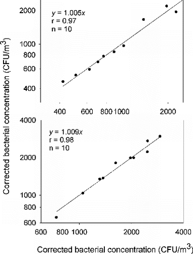
FIG. 7. Electrostatic charge densities of empty dishes, and agar plates before and after sampling.
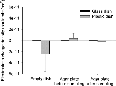
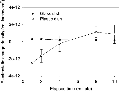
FIG. 8. Decay kinetics of electrostatic charges on dishes as exposed to the atmosphere.