Figures & data
Figure 1. (a) Normalized color ratio (blue band/red band peak height) for laboratory and field samples. The vertical axis in logarithmic scale is for the field samples. (b) The median and 25–75th percentile of the measured color ratio for laboratory samples of known particle mass.
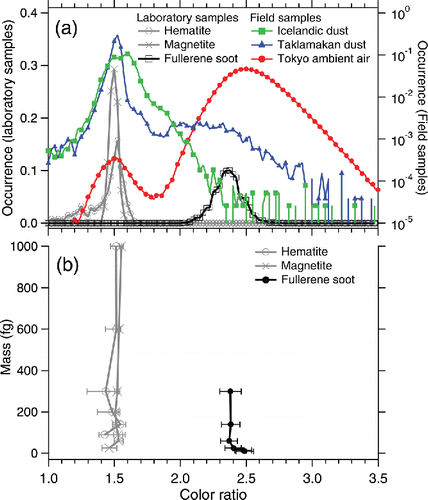
Figure 2. Observed signals related to the incandescence of laboratory samples as a function of particle mass: (a) probability of incandescence in the laser beam of SP2, (b) peak height of the blue-band incandescence signal and associated fitting curves, and (c) timing of the onset of incandescence (toi) relative to the timing at the center of the Gaussian laser beam (tcen). Marker with error bars in panels (a) and (c) denotes the median and 5–95th percentile.
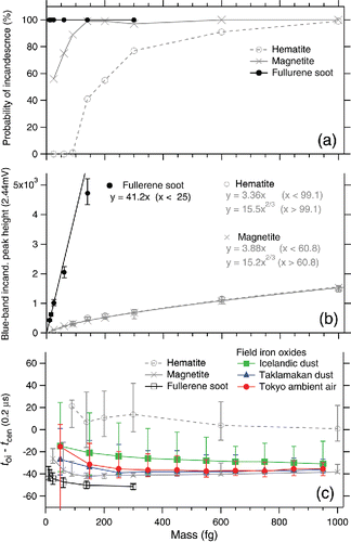
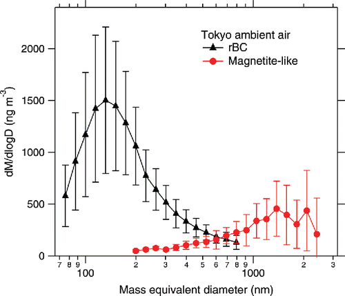
Figure 3. Mass-size distribution of rBC and magnetite-like particles in the Tokyo ambient air. Marker and error bars denote the mean ± standard deviation of 16 values of 30 min averaged data acquired during the 8 h observation period.