
Figures & data
Figure 1. (a) Flight path over Memphis, TN for Filter No. 1 (heaviest loading). The “S” represents initiation of the measurement. (b) Variation of the PSAP absorption coefficient with time (decimal value).
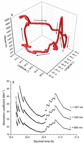
Figure 2. Schematic of the filter transmissivity experimental arrangement.
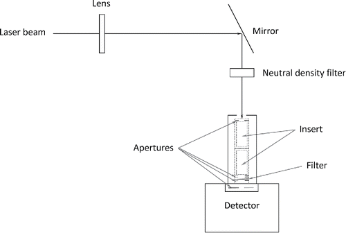
Figure 3. Schematic of the filter absorptivity experimental arrangement.
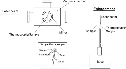
Table 1. Various relationship criteria that must be satisfied to assure proper determination of the particle absorption coefficient.
Figure 4. Magnified images of different PSAP filters under a microscope: (a) blank filter (M = 10) and (b) exposed filter (M = 10). M = microscope magnification. Insert is photograph of a particle-laden PSAP filter. Magnified images of a (c) larger pendent particle hanging from filter fibers (M = 20) and (d) smaller particles loading the filter fiber surface (M = 100).
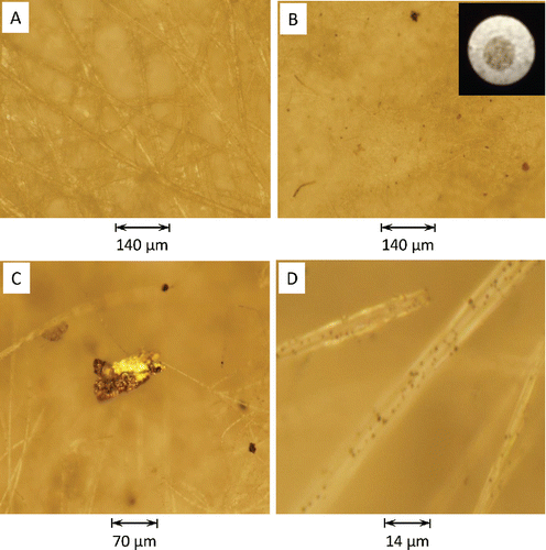
Figure 5. Field-emission scanning electron microscope image of representative particles (for all images: electron beam energy: 10 keV, current: 130 pA, pixel size [nm]: HFW/1024 horizontal pixels [HFW: horizontal field width], secondary-electron mode, and Everhart–Thornley detector). The particle images are arranged in order of decreasing magnification. The magnification (M), and HFW for each respective image are: (a) M: 120 023 x, HFW: 1.07 μm; (b) M: 99 958 x, HFW: 1.28 μm; (c) 50,000 x, HFW: 2.56 μm; (d) M: 20,000 x, HFW: 6.40 μm. Spectra (e) is of a sample particle (see (d)). Sample sits on a germanium substrate. The map was obtained by rastering at 100 µs per pixel for 100×112 pixels per frame and averaging 300 frames. The electron beam energy was set at 20 keV and the current at 0.62 nA.
![Figure 5. Field-emission scanning electron microscope image of representative particles (for all images: electron beam energy: 10 keV, current: 130 pA, pixel size [nm]: HFW/1024 horizontal pixels [HFW: horizontal field width], secondary-electron mode, and Everhart–Thornley detector). The particle images are arranged in order of decreasing magnification. The magnification (M), and HFW for each respective image are: (a) M: 120 023 x, HFW: 1.07 μm; (b) M: 99 958 x, HFW: 1.28 μm; (c) 50,000 x, HFW: 2.56 μm; (d) M: 20,000 x, HFW: 6.40 μm. Spectra (e) is of a sample particle (see (d)). Sample sits on a germanium substrate. The map was obtained by rastering at 100 µs per pixel for 100×112 pixels per frame and averaging 300 frames. The electron beam energy was set at 20 keV and the current at 0.62 nA.](/cms/asset/9802546a-f5dc-49ef-a813-e87d0eb4e8d8/uast_a_1267856_f0005_b.gif)
Table 2. LDTR results for Filter No. 1. Values for the input are the average of two repeated measurements. The subscript H.O.T. refers to higher order terms (for reflections off of the filter internal surfaces) for the transmissivity, τ (Bohren and Huffman Citation1983).(a) Input parameters.
(b) Optical properties and relationship criteria.
(c) Addition relationship criteria.
Table 3. PSAP results for Filter No. 1 (heaviest loading). The given transmissivity, τ, is the value at the cumulated time, tc [s]. The area of the exposed portion for each filter was estimated to be (1.964 ± 0.014) × 10−5 m2.(a) Input parameters.
(b) Optical properties.
(c) Fitting parameters for τp = a1 + a2 e−a3λ.
Figure 6. Variation of the absorption coefficient with wavelength for the different PSAP filters (). All four data points for each filter are fit to an exponential decaying function of the form: α= a1 + a2e−a3λ, see .
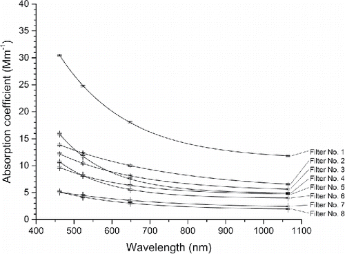
Table 4. Estimation of the particle absorption coefficient, absorption Angström exponent, and enhancement factor from the PSAP and LDTR measurements.
Table 5. Values of the fitting parameters for the exponential decaying function in .