Figures & data
Figure 1. (a) Layout of the test chamber showing core components. (b) Layout of the test chamber showing centerline sampling locations (C1 & C2). (c) Layout of the test chamber showing an overhead view of corner sampling locations (NF: Near-inlet and Far-from-lamp; NN: Near-inlet and Near-lamp; FN: Far-from-inlet and Near-lamp; FF: Far-from-inlet and Far-from-lamp), where the proximity of the pathogen source to each air sampler is shown as dashed (red) lines, and the proximity of the irradiation source to each air sampler is shown as long-dashed (blue) lines. All numerical values are given in units of meters and for the case where all air samplers and both sources were located at 0.80 m above the test chamber floor.
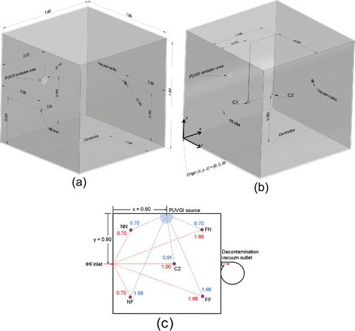
Table 1. Air sampling locations, where increasing x is defined as the downstream direction perpendicular to the Φ6 inlet face, y is the downbeam direction perpendicular to the PUVGI emission face, z is vertically upward from the test chamber floor, and proximity is the minimal linear distance between two locations.
Figure 2. Measured irradiance spectrum at various distances from source.
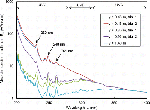
Figure 3. Reduction of recovered pathogen on PTFE filters with increasing PUVGI duration. Recovered pathogen concentration values shown are the mean and standard deviation from triplicate trials (n = 3).
Figure 4. Recovered pathogen mainly found at intermediate particle size in the BC 251 samplers for trials without PUVGI. Recovered pathogen concentration values shown are the mean and standard deviation from duplicate trials (n = 2).
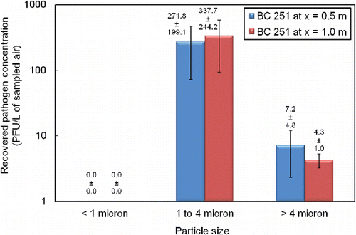
Table 2. Distribution of measured absolute irradiance over various spectral ranges.
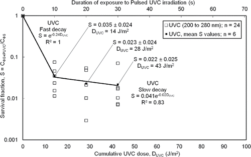
Figure 5. Reduction in survival fraction on PTFE filters with increasing cumulative UVC dose. Survival fraction (S) was calculated as the ratio of the recovered pathogen concentration (for varying PUVC dose) to the recovered pathogen concentration for zero PUVC dose. Cumulative dose (DUVC) was calculated as the product of the exposure duration to PUVC, the pulsing frequency, the pulse width, and the UVC component of absolute irradiance (spectral band from 200 nm to 280 nm). Survival fraction values shown are the mean and standard deviation from triplicate trials at two locations (n = 6).