
Figures & data
Figure 1. Schematic diagram of the PAS. M = mirror, D = dichroic mirror, L = lens, BS = 90:10 beamsplitter, and (A)PD = (avalanche) photodiode.
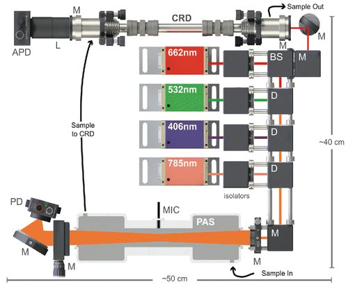
Figure 2. (a) Allan deviation plot of absorption (PAS, solid lines) and extinction (CRD, open circles) data collected with nitrogen gas. (b) Time series and histograms of same data with re-zeroing every 30 min to simulate standard operation of the instruments (30 s average: dark grey lines, 2 min average: light grey (colored) lines, 10 min average: black lines). Dotted lines indicate ±2 standard deviations of the 2-min average data.
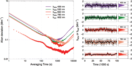
Figure 3. Time series of (a) absorption and extinction from the MultiPAS-IV, (b) absorption from the 7-wavelength aethalometer, and (c) size distribution data from the SMPS (grey (color) scale represents particle number density). The PAS/CRD curves represent 10-min rolling averages of 1-s data; each aethalometer curve represents a 10-min rolling average of 1-min data.
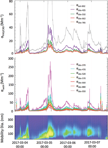
Figure 4. Nigrosin aerosol absorption cross sections measured with the MultiPAS-IV for four different selected mobility diameters (500 nm: diamonds [green], 550 nm: triangles [brown], 600 nm: circles [red], 650 nm: squares [blue]). Curves are Mie theory calculations using the refractive index data of Bluvshtein et al. (Citation2017).
![Figure 4. Nigrosin aerosol absorption cross sections measured with the MultiPAS-IV for four different selected mobility diameters (500 nm: diamonds [green], 550 nm: triangles [brown], 600 nm: circles [red], 650 nm: squares [blue]). Curves are Mie theory calculations using the refractive index data of Bluvshtein et al. (Citation2017).](/cms/asset/42c4759c-6250-4bc7-b841-49df0097b72f/uast_a_1413231_f0004_oc.gif)
Figure 5. Time series and histogram of AAEBC values calculated using the absorbance values at: (a) 662 nm and 785 nm or (b) 532 nm, 662 nm, and 785 nm. Points are sized in relation to the magnitude of absorption at 785 nm. The median values of AAEBC (0.70 and 1.18, respectively) are represented by the dark grey (blue) lines, and the interquartile ranges are represented by the light grey (light blue) shaded regions.
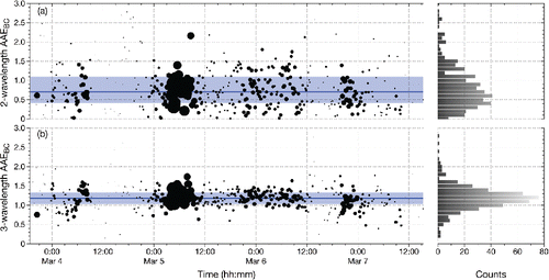
Figure 6. BrC as calculated using the two-wavelength approach to calculating AAEBC. (a) Time series of calculated BrC absorption at 406 nm (dark grey [purple] open circles) and 532 nm light grey (green) open circles. (b) Time series and histograms of fraction of absorption due to BrC at 406 nm (dark grey [purple] closed circles) and 532 nm (light grey [green] closed circles). (c) Time series and histogram of BrC AAE ([orange] circles). Horizontal lines represent median values, while shaded regions represent interquartile ranges.
![Figure 6. BrC as calculated using the two-wavelength approach to calculating AAEBC. (a) Time series of calculated BrC absorption at 406 nm (dark grey [purple] open circles) and 532 nm light grey (green) open circles. (b) Time series and histograms of fraction of absorption due to BrC at 406 nm (dark grey [purple] closed circles) and 532 nm (light grey [green] closed circles). (c) Time series and histogram of BrC AAE ([orange] circles). Horizontal lines represent median values, while shaded regions represent interquartile ranges.](/cms/asset/b1a4332e-1544-437f-bce1-1c1da8a0bddd/uast_a_1413231_f0006_oc.gif)