Figures & data
Figure 1Fractional error in the measured extinction coefficient of an aerosol with size parameter x = πDp /λ due to a finite acceptance angle θ acc.
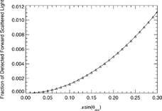
Figure 2Schematics of the LOPES system. The magnified image of the fiber connector jacket shows the side–by–side alignment of the emitting and receiving fibers.
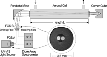
Figure 3Temperature dependence of UV light transmission through the fiber optical system composed of solarization stabilized hydrogen doped silica fibers. The curves show the fiber stabilization during UV light transmission as well as their recovering during dark phases.
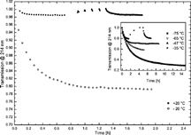
Figure 4Comparison of extinction spectra of Monospher®500 and Monospher®800 quartz glass beads measured by LOPES (solid lines) and calculated by Mie theory (dashed lines). The shaded area gives the 1–σ standard deviation due to instrument noise and aerosol dilution during 5 min averaging time. Narrow particle size distributions of CMD = 520 nm, σ = 1.05, and CMD = 780 nm, σ = 1.03 are necessary to produce the small scale “ripple structure” clearly visible in the measured spectra. Discrepancies in the exact wavelength position of the ripples and in near–infrared spectral slope are attributed to the presence of small agglomerates.
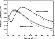
Figure 5Experimental verification of the LOPES spectral performance in the visible by comparison of LOPES extinction coefficients with scattering coefficients of a three–color integrating nephelometer at λ = 450 nm (squares), λ = 550 nm (circles), and λ = 700 nm (triangles). Nonabsorbing spherical quartz glass beads (SSA = 1) of different sizes were used as challenge aerosols. Perfect agreement between LOPES and the nephelometer is indicated the by SSA = 1 (open symbols). For reference, we also report the apparent SSA (solid symbols) based on nephelometer values, which are not corrected for angular truncation and non–Lambertian light source.
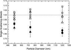
Figure 6Comparison between absorption coefficients from the DM and the PAS. Dotted lines: upper and lower limits of babs DM due to uncertainties in the nephelometer correction (for smoke aerosol).

Figure 7Same as , but for the MAAP instead of the PAS.
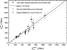
Figure 8Experimental setup for the corn stems burning experiments.
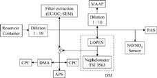
Figure 9SEM image of smoke particles from corn stems smoke.
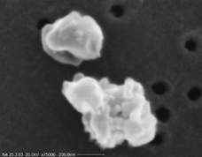
Figure 10Time series of extinction spectra measured with LOPES. The squares represent the result of a Mie calculation based on the measured DMPS size distribution and CNC number concentration. A wavelength–dependent refractive index of m (450 nm) = 1.5 + 0.09i, m (550 nm) = 1.5 + 0.07i, and m (700 nm) = 1.5 + 0.05i was assumed.
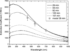
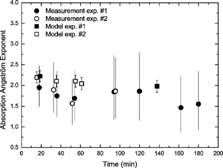
Figure 11Time series of the Ångström exponent deduced from babs DM (circles) and from the modelled absorption cross sections (squares). The error bars of the circles correspond to possible steepest and flattest slopes of babs DM estimated from the highest and lowest value of babs DM (according to the 1–σ standard deviation) at λ = 450 nm and λ = 700 nm.