Figures & data
Table I. Lung abnormalities by score in HRCT scans.
Table II. Lung reactions by grade detected in plain radiographs after radiotherapy.
Figure 1. Radiological pulmonary findings (grade 0–4) at 3, 6 and 12 months (mo) after radiotherapy for the patients evincing some reaction at 12 months.
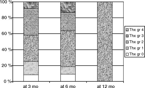
Figure 2. (a) Mean dose volume histograms (DVHs) for all patients and patients with grade 4 pulmonary findings in plain radiographs at either 3 or 6 months after radiation therapy according to type of operation (BCS = breast conserving surgery), M = mastectomy), and (b) DVHs of four individual patients with grade 4 findings also showing pulmonary symptoms.
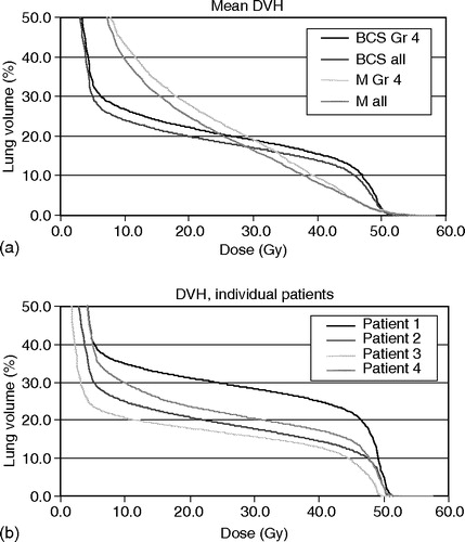
Figure 3. A 57-year old woman (Patient 4) 3 months after radiotherapy of the left breast. Grade-4 lesions = dense uniform opacification in upper lobe and lingula.
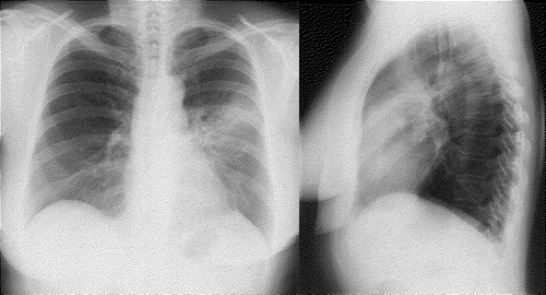
Table III. Lung abnormalities in plain radiographs and in HRCTscans.