Figures & data
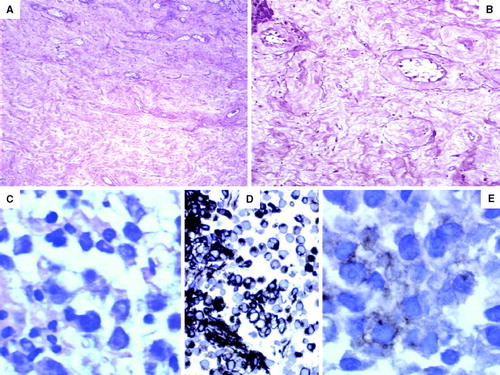
Figure 1. (A) The testis showed severe tubular atrophy, Leydig cell hyperplasia and absence of germ cells (hematoxylin and eosin×200 magnification). (B) Eosinophilic and mixoid transformation with rare hyalinized tubules (elastic tissue stain×200 magnification). (C) Lymph node core biopsy showed uniform cells with clear cytoplasm and clumped chromatin pattern of nucleus. (D) The neoplastic cells showed strong expression of vimentin. (E) Weak expression of placental alckaline phosphatase.