Figures & data
Figure 1. Lymphatic drainage of the prostate (Adapted from [2]).
![Figure 1. Lymphatic drainage of the prostate (Adapted from [2]).](/cms/asset/cdda42a0-9eca-49b0-857c-0e7488834f93/ionc_a_11317377_uf0001_b.jpg)
Figure 2. Extent of lymph node dissection performed: (1) external iliac, (2) obturator fossa, (3) internal iliac (hypogastric), (4) presacral.
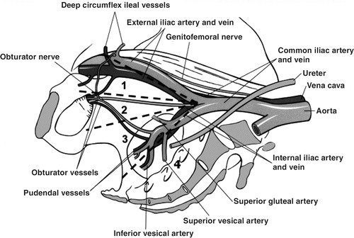
Table I. Distribution of number of nodes and incidence of positive nodes according to the extent of lymph node dissection in retrospective studies.
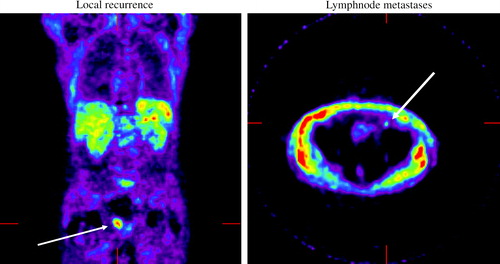
Figure 3. Imaging with PET acetate of local recurrence and lymph node metastases.