Figures & data
Figure 1. Steroid sulfatase detection in MCF-7 cells by immunohistochemistry with anti-steroid sulfatase antibody. (×400).
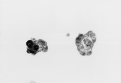
Figure 2. Representative example of steroid sulfatase immunohistochemistry in breast carcinoma. (×200).
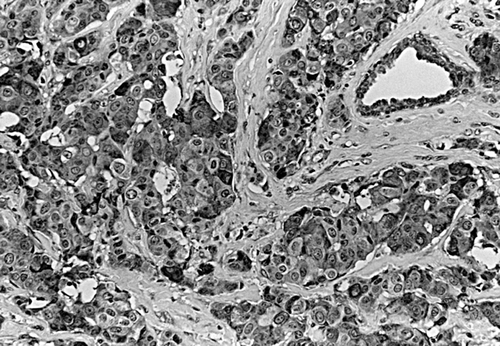
Table I. Association of steroid sulfatase immunoreactivity with clinical parameters.
Table II. Serum estrogens levels in postmenopausal patients with steroid sulfatase positive tumor.
Table III. Change of serum estrogens levels in postmenopausal patients with steroid sulfatase positive tumor before and after operation