Figures & data
Table I. Characteristics of the 4185 patients, University of Florence, 1980–2001.
Figure 1. Cause specific survival for the entire group was 96.9% (±0.2%SE), 93.1% (±0.4%SE) and 83.7 (±0.6%SE) at 3, 5 and 10 respectively.
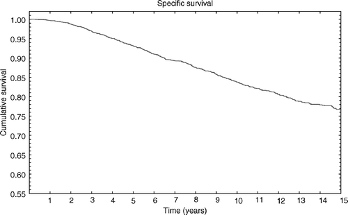
Table II. Site of recurrence in the 224 patients out of the 4185 developing loco-regional recurrence.
Figure 2. The 3, 5 and 10 years actuarial rate for any locoregional recurrence was 2.3% (±0.2%SE), 4.3% (±0.4%SE) and 7.4% (±0.5%SE) respectively.
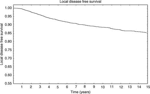
Table III. Correlation between age and LRR.
Figure 3. Local disease free survival by axillary status. 1 = negative axillary nodes; 2 = 1-3 positive axillary nodes; 3 ≥ 3 axillary nodes.
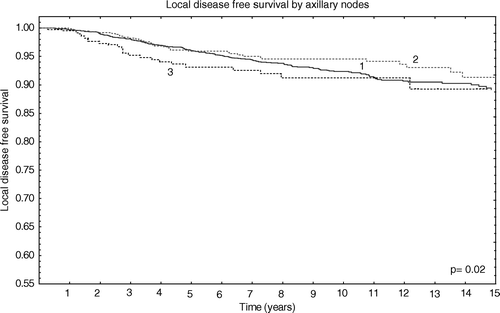
Table IV. Crude and actuarial results for each prognostic factor only in the subgroup of patients with 1 to 3 positive axillary nodes.
Table V. Multivariate analysis for LRR.
Table VI. Multivariate analysis for NR.