Figures & data
Figure 1. Serum (left panel) and tumour (right panel) lactate dehydrogenase activity from either female CDF1 (•) or C3H mice (o) were measured using a colorimetric assay. Measurements were made in mice implanted with differently sized C3H mammary (CDF1 mice) or SCCVII (C3H mice) carcinomas. Individual results are shown, with lines fitted by regression analysis.
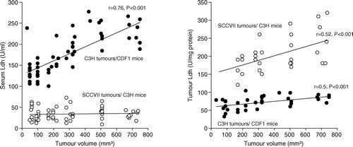
Figure 2. Fold difference in serum (▾) and tumour LDH activity (o), and Ldha mRNA expression (•) in 200 mm3 C3H mammary carcinoma (left panel) and SCCVII carcinoma (right panel) bearing mice treated with 10% oxygen for 1–72 hours compared to controls. Data represent mean ±s.e.m. (n = 10 for each group for serum LDH, n = 4–5 for each group for tumour LDH, n = 3–4 for each group for Ldha mRNA measurement; *p < 0.05, **p < 0.001 compared to control).
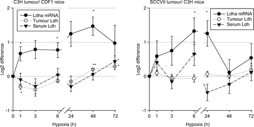
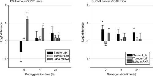
Figure 3. Fold difference in serum (black bars) and tumour LDH activity (gray bars), and Ldha mRNA expression (dark gray bars) in 200 mm3 C3H mammary carcinoma (left panel) and SCCVII carcinoma (right panel) bearing mice treated with 10% oxygen for 12 hours and then reoxygenated for 0, 4 or 24 h when compared to controls. Bars represent means ±s.e.m. (n = 7–9 for each group; *p < 0.05, **p < 0.001 compared to control).