Figures & data
Figure 1. MUltiple Gated Acquisition (MUGA) scan showing left ventricular ejection fraction (LVEF), end-diastolic (ED) diameter and end-systolic (ES) diameter at baseline.
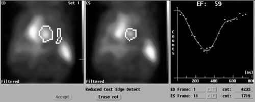
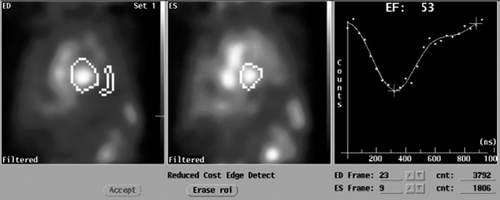
Figure 2. MUltiple Gated Acquisition (MUGA) scan showing left ventricular ejection fraction (LVEF), end-diastolic (ED) diameter and end-systolic (ES) diameter 1 year after the start of sunitinib treatment when cardiac symptoms appeared.
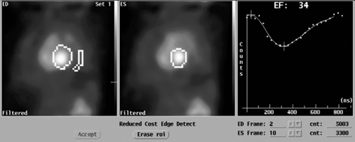
Table I. Results of four tests of MUltiple Gated Acquisition (MUGA) blood pool in a patient with symptoms of heart failure after treatment with sunitinib.
Figure 3. MUltiple Gated Acquisition (MUGA) scan showing left ventricular ejection fraction (LVEF), end-diastolic (ED) diameter and end-systolic (ES) diameter 6 months after the start of therapy for cardiac symptoms.
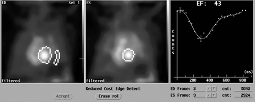
Figure 4. MUltiple Gated Acquisition (MUGA) scan showing left ventricular ejection fraction (LVEF), end-diastolic (ED) diameter and end-systolic (ES) diameter 8 months after the start of therapy for cardiac symptoms.