Figures & data
Figure 1. The structure of oxypeucedanin isolated from the roots of Ostericum koreanum.
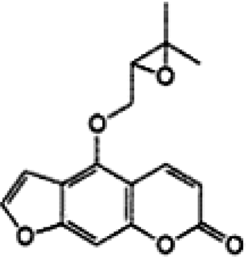
Figure 2. Growth inhibitory effect and cell death-inducing effect of oxypeucedanin in DU145 cells. 1×10⁵ DU 145 cells were plated in 60-mm dishes for 24 h and then treated with DMSO (0.1%, v/v) or oxypeucedanin (25–100 µM). After 24, 48, and 72 h of these treatments, total cells were counted to assess the growth inhibitory effect (A). Total cells were collected and cell viability was determined by trypan blue dye exclusion method (B). The cell growth and cell death data are shown as the mean±SE of three independent plates for each treatment and each sample was counted in duplicate. *, p < 0.05; **, p < 0.001 versus control.
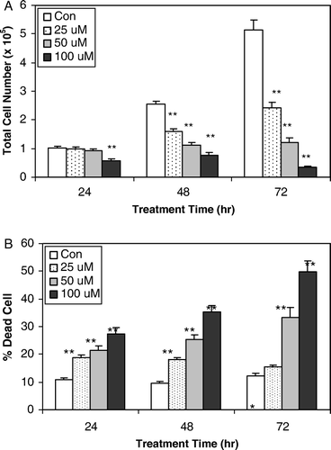
Figure 3. Effects of oxypeucedanin on G2-M cell cycle regulators in DU145 cells. Cells were cultured in RPMI 1640 medium with 10% FBS and treated with DMSO (0.1%, v/v) alone or oxypeucedanin (25–100 µM) for 24 and 48 h. Total cell lysates were then prepared and subjected to SDS-PAGE followed by western blot analysis. Membranes were blotted with anti-Cdc-2, Cyclin A, Cyclin B1, Cdc-25C, Chk1, and Chk2, followed by the appropriate peroxidase-conjugated secondary antibodies, and visualized by ECL detection. Membranes were stripped and reprobed with β-actin antibody as loading control.
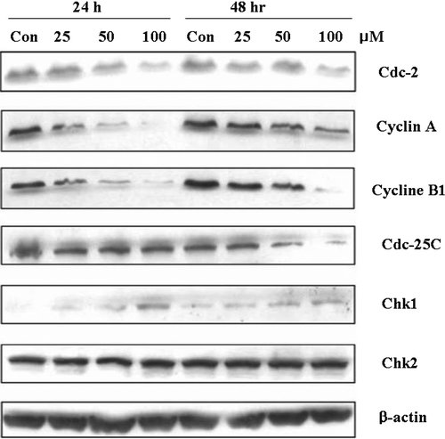
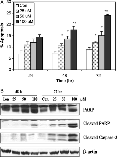
Figure 4. The apoptotic effect of oxypeucedanin on DU145 cells. A. cells were cultured in complete medium and treated with either DMSO vehicle control or 25 to 100 µM oxypeucedanin. After 24, 48, and 72 h, total cells were collected and stained with annexin V/PI followed by flow cytometric analysis. Data are presented as a percentage of annexin V/PI stained cells for each treatment. B. After 48 and 72 h of oxypeucedanin treatment, cell lysates were prepared and SDS-PAGE and western blot analysis were performed for cleaved caspase-3 and total and cleaved PARP using specific antibodies as described in Materials and methods. *, p < 0.05; **, p < 0.001 versus control.