
Figures & data
Figure 1. Unspecified low-grade B-cell lymphoma with features of IgG4-related disease in the resected submandibular gland. (a) Abundant infiltration by small lymphocytic and plasma cells, and features of storiform fibrosis (hematoxylin and eosin staining, magnification ×100). (b) IgG4+ plasma cells located in the periphery of the lymphoma tissue (magnification ×400). (c) IgG + plasma cells (magnification ×400). (d) CD138+ plasma cells (magnification ×400).
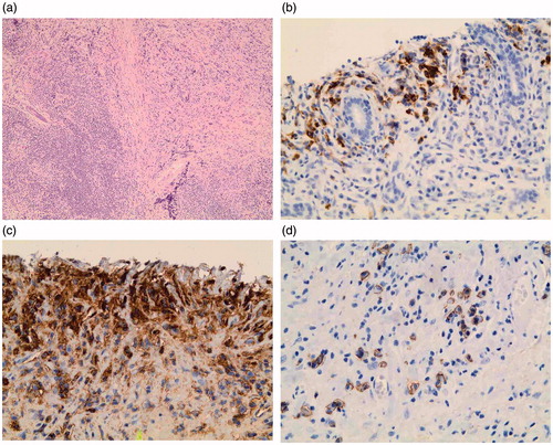
Figure 2. The lung tissue biopsied before lymphoma diagnosis shows dense lymphoplasmacytic infiltration rich in IgG4 + plasma cells, germinal center, and storiform fibrosis (magnification ×100).
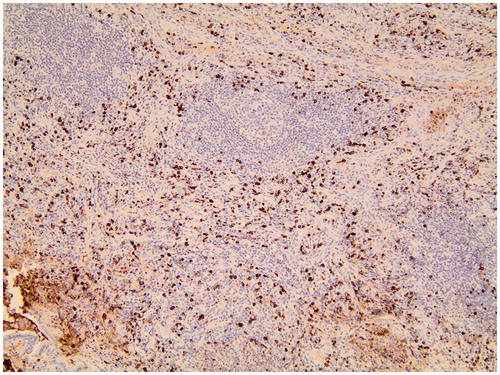
Table 1. IgG4 + plasma cells in the lymphoma tissue of 79 patients with an initial primary Sjögren’s syndrome diagnosis.