Figures & data
Table 1. Patients, tumor characteristics and surgical treatment.
Figure 1. Example dose plan in iPlan (BrainLab) for IMRT of a craniopharyngioma, with coronal, axial and sagittal CT overlaid on MRI with gadolinium (Gd) enhancement. The target volume was defined and treated without setup margins. The eye balls, optic chiasm, nerves and tracts, and brainstem were outlined and defined as organs at risk. The prescription dose of 54 Gy was prescribed to the 90% isodose contour, and the 90% isodose contour was encompassing the target volume.
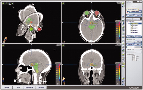
Table 2. Visual function after FSRT.
Table 3. Pituitary hormone deficiency.
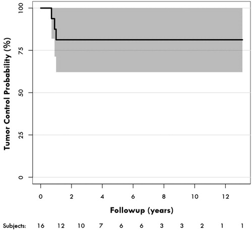
Figure 2. Actuarial tumor control rates at 2, 5 and 10 years after FSRT were 81.3%. Shaded areas represent the 95% confidence intervals.