Figures & data
Table 1. Patient characteristics.
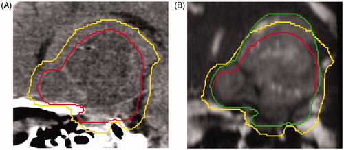
Figure 1. (A) Sagittal view of a computed tomography simulation scan with pretreatment gross tumor volume (GTV; red, inside line) and the 5-mm clinical target volume (CTV) expansion (yellow, outside line). (B) Mid-treatment balanced fast field-echo MRI with the pretreatment GTV (red, inside line), pretreatment 5-mm CTV expansion (yellow, middle line), and mid-treatment GTV contour (green, outside line) extending beyond the pretreatment CTV.