
Figures & data
Table 1. Histopathological tumor characteristics.
Figure 1. (A) Heat map showing graphically the individual recordings of TILs sorted in columns from left to right according to ascending mean stromal TILs values shown in the top row. The rows underneath represents the nine observers recordings arranged from top to bottom according to increasing individual mean values. Single outliers are represented by, e.g., red pixels among otherwise green or yellow pixels or vice versa. (B) A carcinoma with very low mean TILs level (0.03), (C) a carcinoma with a mean TILs level around the 50–60% cut off (0.56) and (D) a carcinoma with a high mean TILs level (0.83). The black bars in (B–D) measures 200 µm (HE, original magnification 100×).
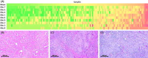
Table 2. Interobserver agreement in assessment of tumor-infiltrating lymphocytes (TILs).