Figures & data
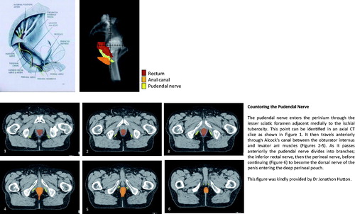
Figure 1. Countoring the pudendal nerve. The pudendal nerve enters the perinium through the lesser sciatic foramen adjacent medially to the ischial tuberosity. This point can be identified in an axial CT slice as shown in (1). It then travels anteriorly through Alcock's canal between the obturator internus and levator ani muscles (2–5). As it passes anteriorily the pudendal nerve divides into branches; the inferior rectal nerve, then the perineal nerve, before continuing (6) to become the dorsal nerve of the penis entering the deep perineal pouch. This figure was kindly provided by Dr Jonathon Hutton