Figures & data
Figure 1. Typical histopathologic findings of ECD and detection of BRAFV600E mutation (case 1). Tissue sample of the perinephric mass is characterized by an extensive xanthogranulomatous fibro-inflammatory reaction. Numerous foamy macrophages are present, accompanied by fibrosis and lymphoplasmacytic cell infiltration (A). Immunohistologically, macrophages were strongly positive for CD68 (B) and Factor XIIIa (C) and negative for CD1a (D) and S100 (E). The BRAFV600E mutation was detected with a BRAFV600E mutation-specific antibody (Clone VE1, Spring Bioscience, Pleasanton, CA) (F). The mutation was confirmed by PCR amplification at amino acid position 600 with an in house protocol and subsequent sequencing using the Pyromark Q24 (QIAGEN, Hilden, Germany, detection limit 10%). Response to treatment assessed by repeated cardiac MRI scanning in case 1. Cardiac MRI scan at presentation (G) and after 6 months (H) and 12 months (I) of SRL treatment, respectively, showed clear regression of pericardial effusion.
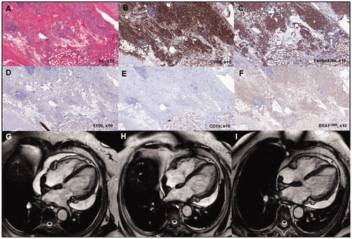
Table 1. Relevant laboratory data of ECD patients on admission.
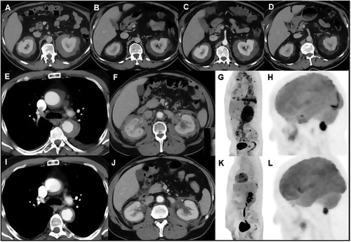
Figure 2. Response to treatment assessed by repeated contrast-enhanced CT scanning in case 2. Abdominal CT scan at presentation (A) and after 6 months of sirolimus (SRL) treatment showed a marked decrease in size and density of the bilateral perirenal soft tissue mass (B). Repeat CT scan 6 months after self-discontinuation of SRL treatment, 8 months after the first follow-up CT scan, showed an increase in size and density of the bilateral perirenal mass (C). Repeat CT scan 6 months after reinstituting SRL treatment again showed a marked decrease in size and density of the bilateral perirenal mass (D). Response to treatment assessed by different imaging modalities in case 3. Thoraco-abdominal CT scan performed at presentation (E,F) and after 6 months of SRL treatment showed a marked decrease in size of the periaortic (I) and perirenal (J) soft tissue mass. 18FDG-PET scan at presentation (G,H) and after 6 months of SRL treatment showed disappearance of 18FDG-uptake at the level of the thoracic (aortic arch) and abdominal aorta, pleuropericardium and bilateral perirenal mass (K) and marked decrease of/disappearance in 18FDG-uptake at the level of the pons and extra-axial lesion near the foramen magna (L), respectively.