
Figures & data
Figure 1. Intense cytoplasmic staining of neoplastic and membranous cells for L1CAM (A) (X100). (B) (X200). (C) Absence of L1CAM staining in normal colonic epithelium. There is L1CAM staining in the ganglionic cells (x200).
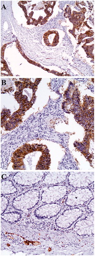
Table 1. Correlation of L1CAM expression with clinicopathological characteristics.
Figure 2. Kaplan–Meier curve depicting progression-free survival (A) and overall survival (B) in months of the study population according to the levels of expression of L1CAM (low vs high). Kaplan–Meier curve illustrating progression-free survival (C) and overall survival (D) of patients with early stage cancer only (stage I and II) and low versus high expression of L1CAM.
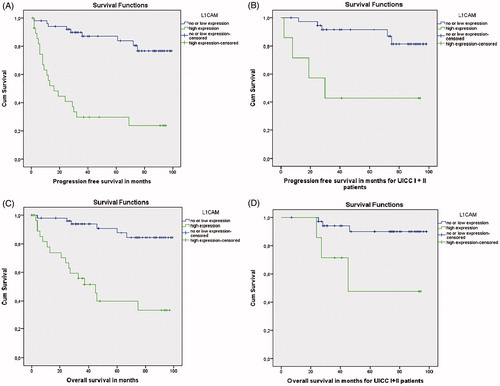
Table 2. Multivariate analysis regarding progression free survival (PFS) and overall survival (OS) using cox proportional hazard models.