
Figures & data
Figure 1. Extension of the CSF within the dural sheaths of the optic nerve (A), trigeminal nerve (B), facial and vestibulo-cochlear nerves (C), the glossopharyngeal, vagus and accessory nerves (D) the hypoglossal nerve (E) and at spinal levels L5/S1 (F) and S1/S2 (G). Images in the left column are acquired using a coronal 3D T2 TSE with a TR/TE = 8000/1094 ms, acquired resolution of 0.75 × 1.5 × 0.75 mm3 and reconstructed to 0.75 × 0.75 × 0.75 mm3 with SPIR fat suppression. The MR images in the right column are acquired using a coronal 3D T2 TSE with TR/TE = 1800/171 ms, acquired resolution of 2.0 × 2.0 × 2.0 mm3 and reconstructed to 1.0 × 1.0 × 1.0 mm3 with SPAIR fat suppression.
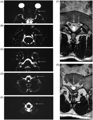
Table 1. CSF extension within the dural sheath of the cranial nerves, measured from the inner table of the skull.