Figures & data
Table 1. Median RBEd (range) across all BSCs for sorted model categories.
Figure 1. The calculated RBEd of the analyzed BSCs across all models for the representative patient. The horizontal solid line shows RBE of 1.1. Open and closed symbols indicate (α/β)x of 2 and 3 Gy, respectively, with exception of CHD, CHE, and WIL. Ent. Cor.: entorhinal cortex; L: left; R: right; LFWM: left frontal white matter; Hippo: hippocampus; SVZ: subventricular zone.
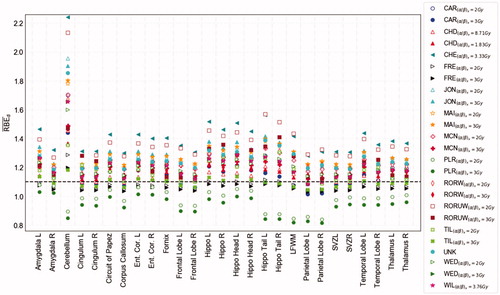
Table 2 . Overview of data point values in experimental databases used to fit the variable RBE models (phenomenological) along with model dependencies.
Figure 2. Minimum and maximum RBEd values for all models (filled green) and the selected set of models (filled yellow) calculated for the patient with target volume closest to the median value. Both input parameters of (α/β)x were applied to the tissue-dependent models. The beige bars show the physical mean dose of each structure. Black dashed line show RBE 1.1, and the two other dashed lines show median values across all structures for all models (green) and the selected set (yellow). Ent. Cor.: entorhinal cortex; L: left; R: right; LFWM: left frontal white matter; Hippo: hippocampus; SVZ: subventricular zone.
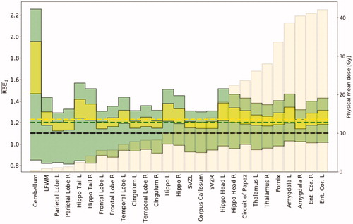
Figure 3. RBEd values of the studied BSCs, arranged in ascending order by the median physical mean dose (beige bars in background) across all patients and both input parameters of (α/β)x. The horizontal dashed lines represent the median, quartiles, and range of the variable RBE models across all BSCs. Ent. Cor.: entorhinal cortex; L: left; R: right; LFWM: left frontal white matter; Hippo: hippocampus; SVZ: subventricular zone.
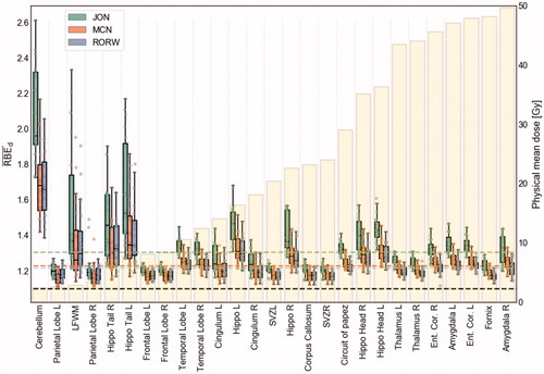
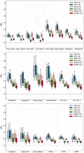
Figure 4. RBEd values for the selected set of models across all patients and (α/β)x of 2 and 3 Gy, in the supratentorial substructures (upper), temporal lobe substructures (middle), and ventricular substructures (lower). Corp.: corpus callosum; Ent. Cor.: entorhinal cortex; Front: frontal; L: left; R: right; Par: parietal; Temp: temporal; SVZ: subventricular zone.