Figures & data
Figure 1. Schematic representation of the three different DIR workflows applied. In WF1, the pMR and its structures were repetitively deformed to the session MR’s. In WF2, the latest MR and structures were deformed to the following session scan without correcting any errors in propagated structures before propagating these to the next session scan. In WF3, the latest MR and structures were deformed to the following session scan with correction of errors in propagated structures before propagating these to the next session scan. Note that the first step in all three workflows is the same.
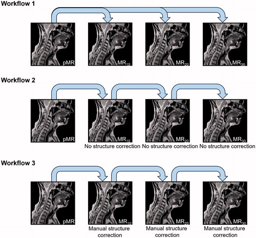
Table 1. (A) Population medians of patient median DSC, MSD and HD calculated for each structure resulting from each of the three deformation workflows as well as the IOV. IOV delineations of the larynx were not performed. (B) Patient subgroup median DSC, MSD and HD of the GTV-N resulting from each of the 3 deformation workflows. Individual deformations were divided into two groups based on whether their volume change was below (relatively small tumour volume change) or above (relatively large tumour volume change) the population median.
Figure 2. 90 percentile surface distance projection images for the propagated right parotid gland structures based on the three workflows as well as for the intra-observer variation.
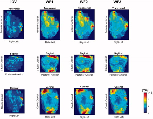
Figure 3. One patient who experienced large relative shrinkage of the GTV-N over the treatment course, as shown by the red curve for manual delineation. The DIR propagated GTV-N from pMR to each session scan in WF1 (green) remains fairly constant in volume compared to the GTV-N propagated from the previous MR, with corrected structures of WF3 (blue), follows the shrinking tumour size better.
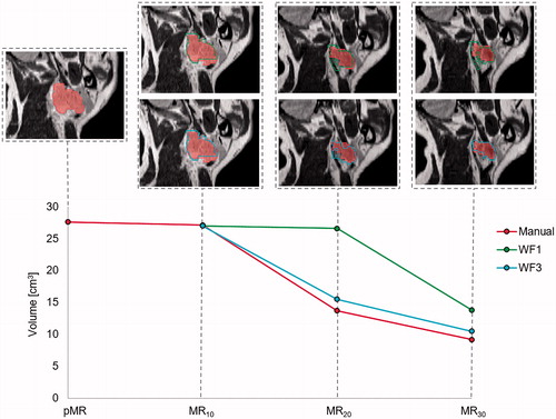
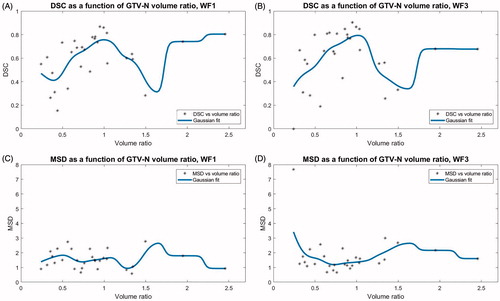
Figure 4. Each patients’ DSC and MSD for WF1 and WF3 plotted against the ratio between the volumes of the GTV-N on the source image for the propagation and the session image, respectively. Volume ratios < 1 indicate GTV-N shrinkage over the treatment course. The running average, shown in blue, was a Gaussian spline function of width 0.1.