
Figures & data
Figure 1. The process of auto-segmentation consists of three steps. (1) A rigid registration between the CT scans in the atlas database and the CT scan to be auto-segmented aiming to obtain the five CT scans with the highest anatomical match. However, for auto-segmentation of the left ventricle wall, only three subjects were used as this resulted in a higher delineation consistency than when using five subjects. (2) The selected CT scans were submitted to the deformable image registration (DIR) algorithm where the suggestion for each structure for the selected patients were transferred to the new CT scan. (3) Finally, the simultaneous truth and performance level estimation (STAPLE) algorithm was used to collapse the multiple delineations into one delineation.
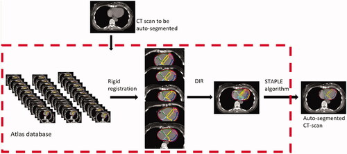
Figure 2. Boxplot of the DSC between the 60 manual delineations (blue dots) performed by 16 observers in CT scans of 12 breast cancer patients and between the automatic and manual delineations (red dots) for the heart and substructures in the final test. Median values for manual and automatic delineations are given at the top, with corresponding p-values for the difference. Abbreviations: Heart: Whole heart; Vent: Ventricle; L: Left; R: Right; Ant: Anterior; Lat: Lateral; Inf: Inferior; Sep: Septal; LMCA: Left Main Coronary Artery; LAD: Left Anterior Descending coronary artery; Cx: Circumflex coronary artery; RCA: Right Coronary Artery; PDA: Posterior Descending Artery; Prox: Proximal; Mid: Middle; Dist: Distal; PT: Pulmonary Trunk; SVC: Superior Vena Cava; IVC: Inferior Vena Cava.
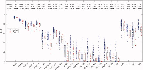
Figure 3. Boxplot of the MSD between the 60 manual delineations, (blue dots) performed by 16 observers in CT scans of 12 breast cancer patients and between the automatic and manual delineations (red dots) for the heart and substructures in the final test. Median values for manual and automatic delineations are given at the top, with corresponding p-values for the difference. Abbreviations: Heart: Whole heart; Vent: Ventricle; L: Left; R: Right; Ant: Anterior; Lat: Lateral; Inf: Inferior; Sep: Septal; LMCA: Left Main Coronary Artery; LAD: Left Anterior Descending coronary artery; Cx: Circumflex coronary artery; RCA: Right Coronary Artery; PDA: Posterior Descending Artery; Prox: Proximal; Mid: Middle; Dist: Distal; PT: Pulmonary Trunk; SVC: Superior Vena Cava; IVC: Inferior Vena Cava.
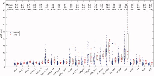
Figure 4. The correlation between RT doses in the automatic delineations (x-axis) and the five manual delineations (y-axis) for the heart and substructures in the final test. The line is x = y (line of identity) where the light gray dots located vertically indicates the variation in the manual delineations. Abbreviations: Heart: Whole heart; Vent: Ventricle; L: Left; R: Right; Ant: Anterior; Lat: Lateral; Inf: Inferior; Sep: Septal; LMCA: Left Main Coronary Artery; LAD: Left Anterior Descending coronary artery; Cx: Circumflex coronary artery; RCA: Right Coronary Artery; PDA: Posterior Descending Artery; Prox: Proximal; Mid: Middle; Dist: Distal; PT: Pulmonary Trunk; SVC: Superior Vena Cava; IVC: Inferior Vena Cava.
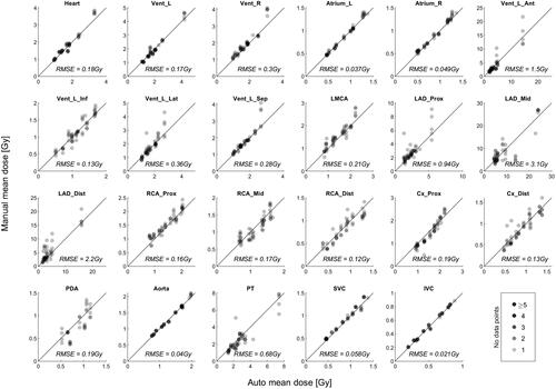
Table 1. Comparison of the method, sample size, and structures for auto-segmentation using atlas-based approach published in the recent years.