
Figures & data
Figure 1. Transaxial co-registered CT and fused images of 18F-FDG PET/CT showing increased tracer uptake in the left upper lung lobe nodule (A and B, arrows), but only weak glucose activity in the lower lobe nodule (C and D, arrows); maximum intensity projection image (E) showing no other sites of abnormal 18F-FDG uptake. At subsequent 11C-methionine PET/CT, transaxial co-registered CT and fused images showed increased and similar uptake of amino acidic tracer both in the left upper lung lobe nodule (F and G, arrows) and in the lower lobe nodule (H and I, arrows); maximum intensity projection image (L) showing no other sites of abnormal 11C-methionine uptake, so suggesting ACC oligo-metastatic lung involvement.
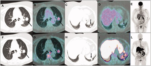
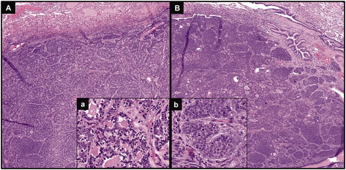
Figure 2. Hematoxylin & eosin staining of both lung nodules confirming metastases from the previous ACC, with basaloid neoplastic cells arranged in a predominantly solid architecture (A and B, original magnification 40×), presenting some cribriform areas, with glandular structures containing amorphous material (inset a; original magnification 200×) and some mitotic features (inset b; original magnification 400×).
Data availability statement
Due to the nature of this research (case report), the data are not publicly available since containing personal information that could compromise the privacy of the patient.