
Figures & data
Figure 1. Rigid co-registration of the staging- and relapse scans for two patients (a–e and f–h). (a) Staging PET/CT-scan. Arrow pointing to an initially involved PET positive supraclavicular lymph node. (b) CT part of the staging PET/CT-scan. Delineation of the PET positive (cyan) and the anatomically involved volume (red). (c) Relapse PET/CT-scan. Arrow pointing to a PET positive relapsed supraclavicular lymph node. (d) CT part of the relapse PET/CT-scan. Delineation of the PET positive (yellow) and the anatomically involved volume (blue). (e) Fusion of the staging- and the relapse PET/CT-scans. The relapse is in the exact same lymph node as initially involved. (f) CT part of the staging PET/CT-scan. Delineation of a PET positive (cyan) and two anatomically involved mediastinal lymph nodes (red). (g) CT part of the relapse PET/CT-scan. Delineation of a PET positive (yellow) and anatomically involved (blue) mediastinal lymph node. (h) Fusion of the staging- and the relapse scans. The relapse is in the exact same lymph node as initially involved.CT: computed tomography; PET: positron emission tomography.
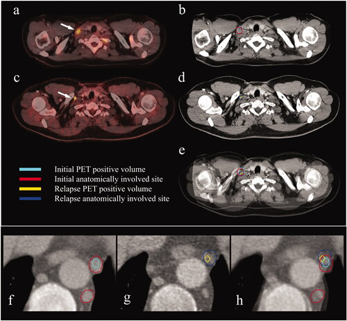
Table 1. Details of relapse locations.
Table 2. Baseline characteristics.
Table 3. Treatment characteristics.
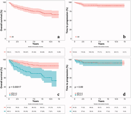
Figure 2. Overall survival (a) and time to progression (b) for all patients. Overall survival (c) and time to progression (d) stratified on IPS groups. Four patients could not be stratified to a group due to missing values in the IPS and were left out of the stratified analyses. IPS: International Prognostic Score.
Supplemental Material
Download PDF (289.8 KB)Supplemental Material
Download PDF (82.6 KB)Data availability statement
The data generated during the current study are not publicly available due to regulations on personal data protection but are available from the corresponding author on reasonable request.