
Figures & data
Table 1. Clinical and prognostic factors in relation to histotype.
Figure 1. Representative microscope (x20) photographs of immunohistochemical immune cell markers (CD8, cytotoxic T-cells; CD3, peripheral T-cells; CD20, precursor and mature B-cells; FOXP3, regulatory T-cells; CD163, macrophage lineage) high expression in leiomyosarcomas (LMS).
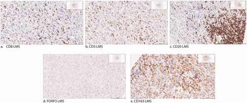
Table 2. Spearman’s correlation between immune cell markers.
Table 3. Correlation between immune cell markers and known prognostic factors.
Table 4. Metastasis-free and Overall Survival. Univariable and multivariable analyses.
Supplemental material
Supplemental Material
Download MS Word (15 KB)Data availability statement
The data that support the findings of this study are available from the corresponding author upon reasonable request.