
Figures & data
Figure 1. Profiles of APOE transcription (A-B), methylation (C-D) in paracancerous and HCC tissues. LIHC stands for liver hepatocellular carcinoma.
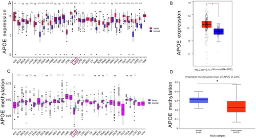
Figure 2. Expression profiles of APOE transcription in GEO, TCGA and ICGC datasets (A-I), and meta-analysis of APOE expression (J). TCGA stands for The Cancer Genome Atlas, GEO stands for Gene Expression Omnibus, ICGC stands for International Cancer Genome Consortium.
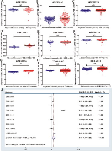
Figure 3. APOE methylation is associated with immune infiltration and immune activation in HCC. (A) Heatmap displayed APOE methylation associated relative abundance of 28 immune cells in HCC. (B) The relationship between the methylation of APOE and 28 immune cells in HCC. (C) The comparison of TILCs in APOE hypermethylation and hypomethylation subgroups. (D) Heatmap showing a relative association between APOE methylation and 26 immunity-related gene sets. (E)The relationship between 26 immunity-related gene sets and APOE methylation in HCC
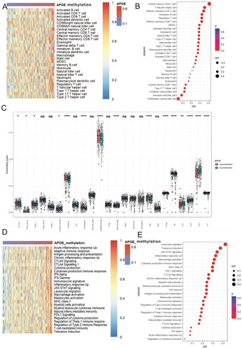
Figure 4. APOE methylation is associated with the steps of the cancer immunity cycle (A-B) and T-cell inflammatory signature (TIS) scores of HCC (C).

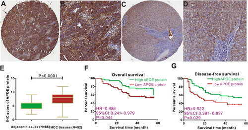
Figure 5. Expression and survival significance of APOE protein in HCC patients. Representative figures of APOE staining in HCC tissues (A-B) and adjacent liver tissues (C-D). APOE protein is highly expressed in HCC tissues (E). HCC patients with APOE over-expression displayed favorable overall survival (F) and disease-free survival (G) than those with low APOE expression.
Supplemental Material
Download MS Word (82.5 MB)Data availability statement
All data generated or analyzed during this study are included in this article and its supplementary information files.