Figures & data
Figure 1. Schematic representation of the initiation of the angiogenic cascade in a cell. Whenever sufficient oxygen is supplied to the cell (upper half of figure), HIF-1α is metabolized in the cytoplasm, and the angiogenic cascade is not started. Whenever insufficient oxygen is supplied to the cell (lower half of the picture), HIF-1α proceeds to the cell nucleus and binds to HIF-1β. The HIF-1α-HIF-1β-complex binds to the HRE, and angiogenic factor transcription is initiated.
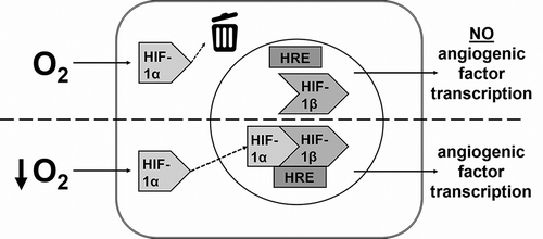
Table 1. Primer sequences.
Table 2. Table indicating p values of differences in cell proliferation of 0.1, 1, and 21% oxygen as compared to all other tested oxygen percentages, for five time points.
Figure 2. Line graph representing the number of cells per well at several oxygen percentages measured at five time points over 14 days. Error bars represent the standard error of the mean.
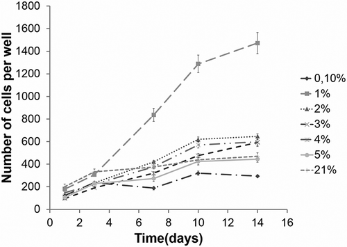
Table 3. P values of differences in CCMR of 21% oxygen as compared to all other tested oxygen percentages, for five time points.
Figure 3. Line graph representing the corrected cell metabolic rate. This was calculated at five time points over 14 days for several oxygen percentages. Error bars represent the standard error of the mean.
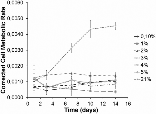
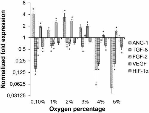
Figure 4. Bar chart representing the normalized fold expression of ANG-1, TGF-β, FGF-2, VEGF, and HIF-1α at several oxygen percentages when compared to expression of these factors at 21% oxygen. Normalized fold expressions that differ significantly from normalized fold expression at 21% oxygen are indicated with an asterisk. Error bars represent the standard error of the mean.