Figures & data
Figure 1. Picture of a tibialis anterior muscle’s tendon from a WT (left panel) and a Col1a1Jrt/+ mice (right panel). Note the difference in color and the gluey aspect of the Col1a1Jrt/+ mouse’s tendon.
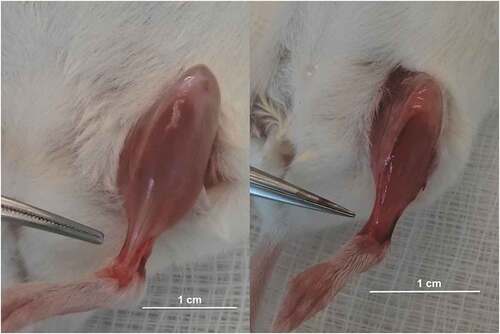
Figure 2. Anatomical and compositional properties of the FDL tendons in WT (black bars; n= 10) and Col1A1Jrt mice (white bars; n = 10). Results are presented as mean and SEM. Panel A. Tendon width; Panel B. Tendon thickness; Panel C. Tendon cross-sectional area; Panel D. Collagen content relative to dry weight. *** indicates significance at the <0.001 level, ns: non-significant at >0.05.
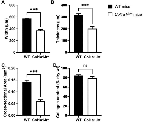
Figure 3. Mechanical properties of the FDL tendons in WT (black bars; n=10) and Col1A1Jrt mice (white bars; n=10). Results are presented as mean and SEM. Panel A. Tendon peak force; Panel B. Tendon yield force; Panel C. Peak stiffness. *** indicates significance at the <0.001 level.
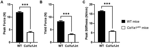
Figure 4. Material properties of the FDL tendons in WT (black bars; n=10) and Col1A1Jrt mice (white bars; n=10). Results are presented as mean and SEM. Panel A. Tendon peak stress; Panel B. Tendon yield stress; Panel C. Tendon Young’s modulus. Panel D. Relative stress-relaxation at 60 s. ** indicates significance at the <0.01 level, ns: non-significant at >0.05.
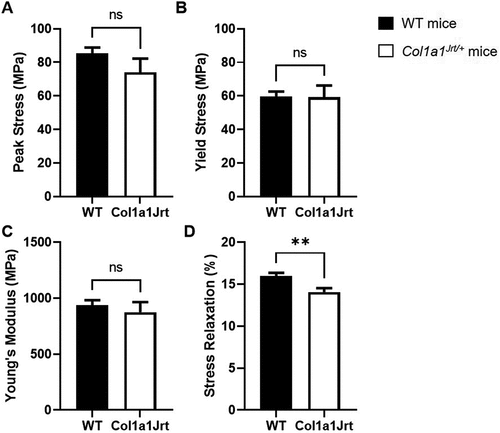
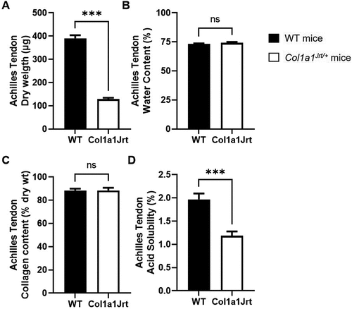
Figure 5. Composition and solubility of the Achilles tendon in WT (black bars; n = 10) and Col1A1Jrt mice (white bars; n = 10). Results are presented as mean and SEM. Panel A. Tendon dry weight; Panel B. Tendon water content; Panel C. Collagen content relative to dry weight; Panel D. Collagen solubility in acetic acid as a percent of total collagen. *** indicates significance at the <0.001 level, ns: non-significant at >0.05.