Figures & data
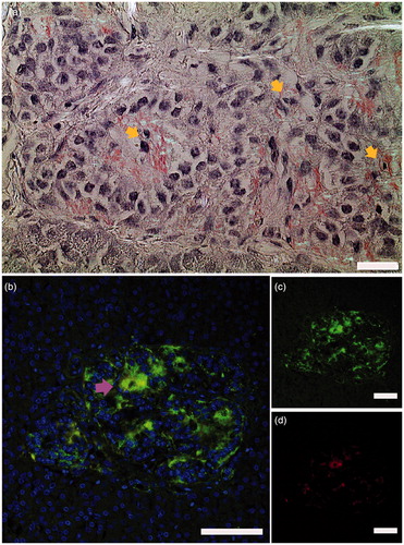
Figure 1. Islet amyloid was detected in pancreata obtained from patients newly diagnosed with type 1 diabetes. (a) a section from a pancreas tail biopsy stained with Congo red showing multiple islets with amyloid. Intracellular amyloid is associated with pyknotic cell nuclei indicated with yellow arrows. (b–d) IAPP was detected with a hIAPP specific antibody and visualized with an Alexa-488-labelled detection antibody (green), and amyloid by a subsequent staining with Congo red (red). Co-localization of IAPP immunoreactivity and amyloid (yellow) indicated by arrowhead in magenta ensures that amyloid is made up of IAPP. Bar 20 µm.