
Figures & data
Figure 1. Monoclonal antibody staining for cytokeratin 19 in large-core needle biopsy of breast carcinoma obtained for diagnosis. Note the intense diffuse staining at membrane and intracytoplasmic level in 100% of tumor cells.
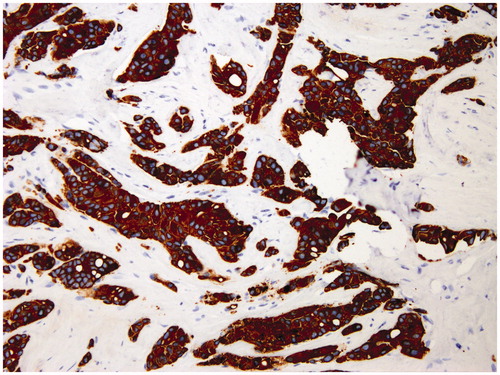
Figure 2. Macrometastasis, micrometastasis, and isolated tumor cells in sentinel lymph nodes of women with breast cancer. (A) H&E section (×40) shows abundant clusters of epithelial cells on a fibrous stroma at an intranodal location. The clusters are larger than 2 mm. (B) H&E section (×100) shows a focus of epithelial cells on a fibrous stroma at an intranodal location measuring more than 0.2 mm but less than 2 mm. (C) H&E section (×100) demonstrates a small cluster of epithelial cells in the subcapsular region. The cluster measures less than 0.2 mm.
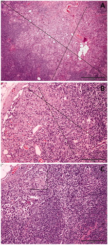
Table 1. Patient and tumor characteristics.
Table 2. Sentinel and axillary lymph node dissection characterization.
Figure 3. Total antioxidant capacity (TAC) (A), thiobarbituric acid reactive substances (TBARS) (B), and carbonyl group content (C) in negative sentinel lymph nodes (SLNs), SLNs with micrometastasis, and SLNs with macrometastasis of women with breast cancer. Results are expressed in nmol of Trolox equivalents per mg of protein for TAC, in ng of malondialdehyde per mg of protein for TBARS, and in nmol per mg of protein for protein carbonyls (mean ± SEM; n = 13–43; *P < 0.05; **P < 0.01; aP < 0.05 micrometastasis versus macrometastasis).
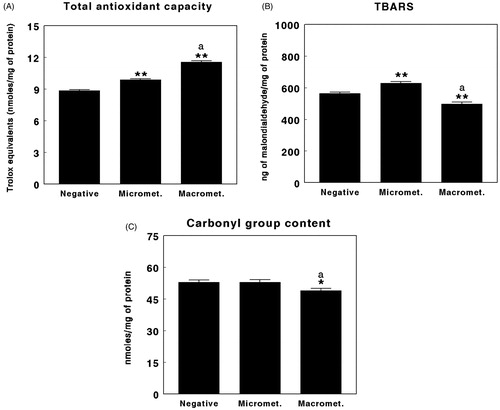
Figure 4. Reduced glutathione (GSH) in negative sentinel lymph nodes (SLNs), SLNs with micrometastasis, and SLNs with macrometastasis of women with breast cancer. Results are expressed in ng per mg of protein (mean ± SEM; n = 13–43; *P < 0.05; aP < 0.05 micrometastasis versus macrometastasis).
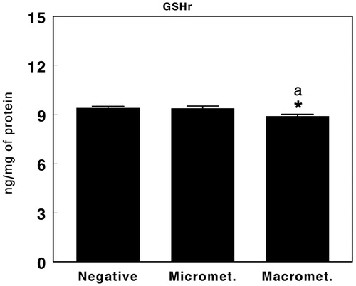
Figure 5. Superoxide dismutase (SOD) (A), catalase (CAT) (B), and glutathione peroxidase (GPx) (C) activity in negative sentinel lymph nodes (SLNs), SLNs with micrometastasis, and SLNs with macrometastasis of women with breast cancer. Results are expressed as percentage inhibition for SOD, in nmol per min and per mg of protein for CAT, and in µmol per min per mg of protein for GPx (mean ± SEM; n = 13–43; **P < 0.01; aP > 0.05 micrometastasis versus macrometastasis).
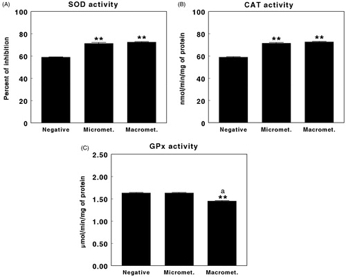
Table 3. Oxidative stress parameters, non-enzyme and enzyme antioxidant defense systems in the sentinel lymph node (SLN) of women with breast cancer.