Figures & data
Table 1. List of antibodies used.
Figure 1. A: SR-PCI of a right human cochlea. Framed area is magnified in B. The saccule (S) and stapes plate (arrow) can be seen. B: Cochlear tissue is detectable, including Reissner’s membrane (RM), SL, and BM. C: SR-PCI of a left human ear at the level of the RW. The RM, BM, SL, and round window membrane (RWM) are clearly visible, as well as the limbus spirale. The SL facing the ST shows increased contrast (* and arrow). D–G: Sections showing the lateral attachment of the RW near the SL. There is some contrast enhancement of the ST wall facing the RW (arrows, SSL). There is often a space (*) between the RWM and the LW. (BC: basilar crest; BM: basilar membrane; LW: lateral wall; OC: otic capsule; OSL: osseous spiral lamina; RM: Reissner’s membrane; RW: round window; RWM: round window membrane; S: saccule; SL: spiral ligament; SR-PCI: synchrotron radiation phase contrast imaging; SSL: secondary spiral lamina; ST: scala tympani; SV: scala vestibuli).
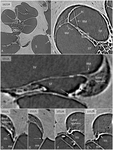
Figure 2. Serial SR-PCI sections from two right ears (A1–4; B1–4) at the cul-de-sac (*) of the ST space (*). The RWM extends basally beyond the level of the BM, and they do not seem to unite. The SSL separates the RWM from the BM (B3, 4) and the SL (A2–4). (For abbreviations, see legend to ).
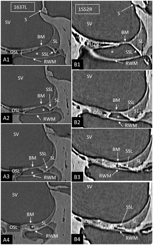
Figure 3. A: SR-PCI section of a left ear showing RM, SL (arrows), and RWM at the cul-de-sac of the endolymphatic space. There is an increased contrast (arrows) of the inferior region of the SL facing the ST. B: SV view of the 3D reconstructed tissues in the same cochlea. The basal end of the BM is seen together with the SL (blue) and OSL (yellow). C: Slightly angled view demonstrates the SSL (arrow). D: Postero-inferior view shows the BM in the SV and the external surface of the RWM with surrounding SSL. E: Infero-lateral view of the basal end of the BM where it joins with the SSL (*), SL, and OSL (encircled). (For abbreviations, see legend to ).
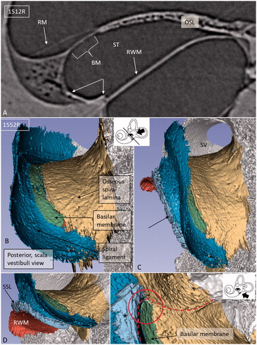
Figure 4. Different angular views of the 3D reconstructed RWM and neighboring soft tissues in a left human ear. A: Infero-medial view shows the relationship between the BM and the SSL (*). B: Same ear viewed from the middle ear displays the relationship between the RWM and stapes. The posterior portion of the RWM lies almost horizontal. C: Infero-medial view of the basal end of the BM and the RWM. Framed area is magnified in F (* = SSL). D: Lateral view of the SL (dark blue) (* = SSL). E: Infero-lateral view with conical shape. F: Magnified framed area in C. The BM is separated from the RWM (arrow). G: Infero-lateral view of the RWM and SL (blue). The close relationship between the SL and RWM is seen. H: Same view as G after removal of RWM (delineated). Inset shows a single SR-PCI section of the medial wall of the round window niche (RWN) (*) and the OSL (yellow). (For abbreviations, see legend to ).
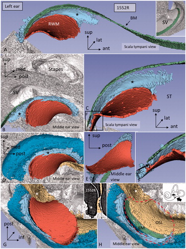
Figure 5. Confocal immunohistochemistry of the human cochlea at the level of the RW. Upper image: The RWM expresses elastin. Some elastic fibers radiate between the BM and the SSL. Lower image: The SSL expresses type II collagen. The OSL also expresses type II collagen. (For abbreviations, see legend to ).
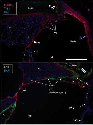
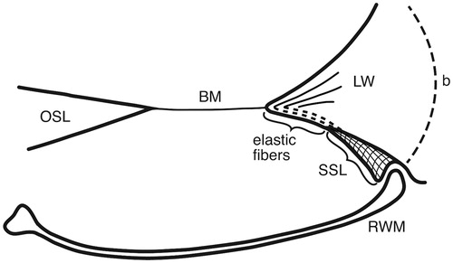
Figure 6. Drawing showing the principal arrangement of the BM and its attachment to the LW at the RWM in a human cochlea. The BM contains radial fibers which reach the BC and radiate into the SL. Fibers express the elastin path between the BC and the SSL. (b: bone; for other abbreviations, see legend to ).