Figures & data
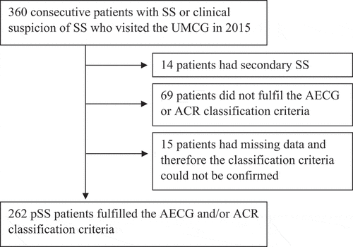
Figure 1. Flowchart of primary Sjögren’s syndrome (pSS) patients included in the analyses. SS, Sjögren’s syndrome; UMCG, University Medical Center Groningen.

Table 1. Number and types of pulmonary manifestations which are possibly or assumed to be related to primary Sjögren’s syndrome (pSS).
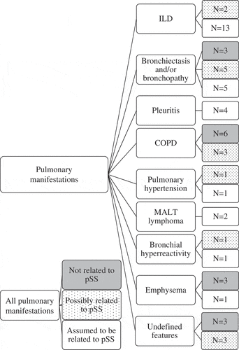
Figure 2. Flowchart of pulmonary manifestations related and not related to primary Sjögren’s syndrome (pSS): all manifestations (26% of the pSS patients), not related to pSS (11%), possibly related to pSS (5%), and assumed to be related to pSS (10%). COPD, chronic obstructive pulmonary disease; ILD, interstitial lung disease; MALT, mucosa-associated lymphoid tissue.

Figure 3. Types of interstitial pneumonia on high-resolution computed tomography (HRCT). (A) A primary Sjögren’s syndrome (pSS) patient with fibrotic non-specific interstitial pneumonia. HRCT shows reticular abnormalities with traction bronchiolectasis (white arrow), ground-glass opacity and lymphadenopathy. (B) A pSS patient with lymphocytic interstitial pneumonia. HRCT shows multiple air cysts (white arrowheads), ground-glass opacity, and traction bronchiolectasis (white arrow). (C) A pSS patient with organizing pneumonia. HRCT shows bilateral patchy consolidations with peripheral and peribronchial predominance and an air cyst (white arrowhead).

Table 2. Pulmonary diagnostics.
Table 3. Characteristics of primary Sjögren’s syndrome (pSS) patients with and without pulmonary involvement and with active pulmonary involvement according to the pulmonary domain of the EULAR Sjögren’s Syndrome Disease Activity Index (ESSDAI).
