
Figures & data
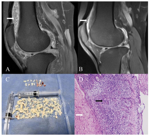
Figure 1. Magnetic resonance imaging, specimens, and pathological findings in the case. (A) Fat-suppressed T2-weighted imaging (T2WI) showing numerous ‘watermelon seed’-like rice bodies within the knee joint cavity, presenting as hypointense compared to joint effusion, with synovial proliferation. (B) Fat-suppressed T2WI showing a marked reduction in synovial proliferation. (C) Photograph of a large number of rice bodies retrieved from arthroscopic lavage. (D) Extensive fibrin exudation and necrotic degenerated tissue (white arrows), with focal formation of inflammatory granulomas (black arrows) (H&E, × 200).
Data availability statement
Data are available from the corresponding author upon reasonable request.