643
Views
6
CrossRef citations to date
0
Altmetric
Articles

A new epizoic laelapid mite from the New Zealand sand scarab Pericoptus truncatus larvae and its isotopic ecology

&
Pages 187-199 | Received 03 Aug 2011, Accepted 28 Sep 2011, Published online: 21 Sep 2012

Figures & data

Figure 1 Mumulaelaps ammochostos sp. n. Female. Dorsal shield setae; legs show location of macrosetae with other setae removed except al1 and pl1 as spines. Idiosomal setal names follow Lindquist & Evans (Citation1965) and leg setal names follow Evans (Citation1963).
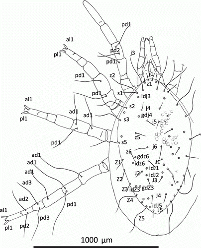
Figure 1  Mumulaelaps ammochostos sp. n. Female. Dorsal shield setae; legs show location of macrosetae with other setae removed except al1 and pl1 as spines. Idiosomal setal names follow Lindquist & Evans (Citation1965) and leg setal names follow Evans (Citation1963).

Figures 2–6 Mumulaelaps ammochostos sp. n. Female. 2, Hypostome, sternal and epigynal shield. 3, Dorsal view of fimbriate internal mala and labrum with chelicera removed. 4, Chelicerae. 5, Venter of mite showing lateral reduction in sternum and reduction in endopodal shields. 6, Lateral view of mite with peritreme and long post lateral setae. Idiosomal setal names follow Lindquist & Evans (Citation1965).
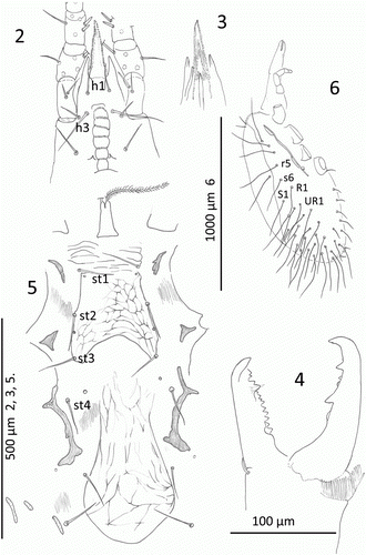
Figures 2–6  Mumulaelaps ammochostos sp. n. Female. 2, Hypostome, sternal and epigynal shield. 3, Dorsal view of fimbriate internal mala and labrum with chelicera removed. 4, Chelicerae. 5, Venter of mite showing lateral reduction in sternum and reduction in endopodal shields. 6, Lateral view of mite with peritreme and long post lateral setae. Idiosomal setal names follow Lindquist & Evans (Citation1965).

Figures 7–15 Mumulaelaps ammochostos sp. n. Ventral views of legs. 711, Female legs. 7, Leg I, genua and tibia. 8, Leg II, genua, tibia and tarsus. 9, Leg III, genua, tibia and tarsus. 10, Leg IV, genua, tibia and tarsus. 11, Femur II, ventral view. 1215, Male legs. 12, Femur II, ventral view showing stout av1 as sexual dimorphism and as wider setae al1 and pl1 as flattened blunt spines. 13, Leg II. 14, Leg III. 15, Leg IV. Leg setal names follow Evans (Citation1963).
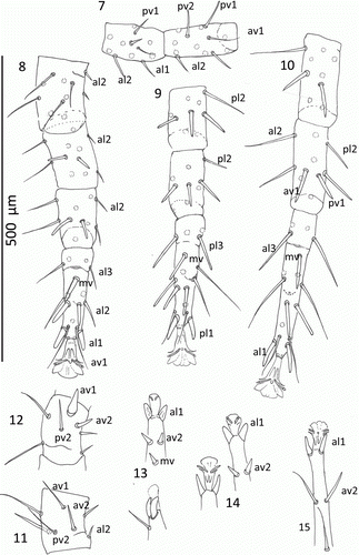
Figures 7–15  Mumulaelaps ammochostos sp. n. Ventral views of legs. 7–11, Female legs. 7, Leg I, genua and tibia. 8, Leg II, genua, tibia and tarsus. 9, Leg III, genua, tibia and tarsus. 10, Leg IV, genua, tibia and tarsus. 11, Femur II, ventral view. 12–15, Male legs. 12, Femur II, ventral view showing stout av1 as sexual dimorphism and as wider setae al1 and pl1 as flattened blunt spines. 13, Leg II. 14, Leg III. 15, Leg IV. Leg setal names follow Evans (Citation1963).

Figures 16–18 Mumulaelaps ammochostos sp. n. Male. 16, Ventral view of sterniventral shield with asymmetry and islands of striate cuticle within the shield. 17, Chelicera and spermatodactyl, axial view. 18, Chelicerae and spermatodactyl, antaxial view. Idiosomal setal names follow Lindquist & Evans (Citation1965).
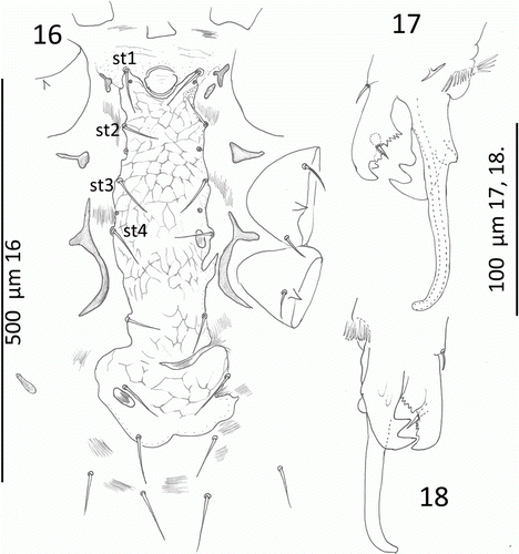
Figures 16–18  Mumulaelaps ammochostos sp. n. Male. 16, Ventral view of sterniventral shield with asymmetry and islands of striate cuticle within the shield. 17, Chelicera and spermatodactyl, axial view. 18, Chelicerae and spermatodactyl, antaxial view. Idiosomal setal names follow Lindquist & Evans (Citation1965).

Figure 19 Isotope data (mean±SEM) for Mumulaelaps ammochostos sp. n. and contextual material. Larval head capsule (n=7 samples) and fat body (n=7) data represent chitinaceous and lipid material respectively for Pericoptustruncatus. Drift wood samples (n=4) came from material supporting the P. truncatus; nitrogen content was too low for δ15N analysis, and is arbitrarily set to zero for display purposes. Mites were too small for individual analysis and so were analysed as four replicates of 3–7 pooled individuals.
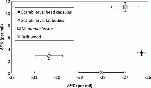
Figure 19  Isotope data (mean±SEM) for Mumulaelaps ammochostos sp. n. and contextual material. Larval head capsule (n=7 samples) and fat body (n=7) data represent chitinaceous and lipid material respectively for Pericoptus truncatus. Drift wood samples (n=4) came from material supporting the P. truncatus; nitrogen content was too low for δ15N analysis, and is arbitrarily set to zero for display purposes. Mites were too small for individual analysis and so were analysed as four replicates of 3–7 pooled individuals.

Reprints and Corporate Permissions

Please note: Selecting permissions does not provide access to the full text of the article, please see our help page How do I view content?

To request a reprint or corporate permissions for this article, please click on the relevant link below:

Academic Permissions

Please note: Selecting permissions does not provide access to the full text of the article, please see our help page How do I view content?

Obtain permissions instantly via Rightslink by clicking on the button below:

If you are unable to obtain permissions via Rightslink, please complete and submit this Permissions form. For more information, please visit our Permissions help page.