339
Views
6
CrossRef citations to date
0
Altmetric
Research articles

Redescription of Tubulanus mawsoni (Wheeler 1940) comb. nov. (Palaeonemertea: Tubulanidae) from the Bellingshausen Sea (Antarctica)

&
Pages 263-279 | Received 31 Oct 2012, Accepted 07 Jan 2013, Published online: 24 Apr 2013

Figures & data

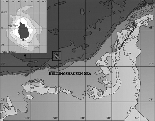
Figure 1 Position of station 5 during Bentart-03 survey, where Tubulanus mawsoni (Wheeler, 1940) comb. nov. was found.
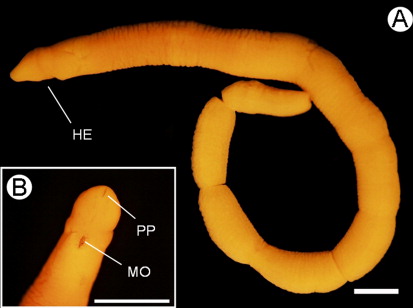
Figure 2 Tubulanus mawsoni (Wheeler, Citation1940) comb. nov.A, Immature female specimen after fixation in Bouin's fluid. B, Detail of the head in ventral view. HE, head; MO, mouth; PP, proboscis pore. Scale bars: 1 mm.
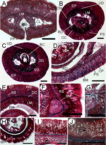
Figure 3 Tubulanus mawsoni (Wheeler, Citation1940) comb. nov.A, Transverse section at the proboscis pore showing the dorsoventral fibres in cerebral lacunae. B, Transverse section through the posterior rhynchodaeal region showing the glandular cells shared by the epidermis and the rhynchodaeal epithelia. C, Transverse section through the cerebral region at level of the proboscis insertion. D, Transverse section through the rhynchodaeal region showing the connection between rhynchodaeal and epidermal serous glands (arrowhead). E, Transverse section through the buccal region; note two different types of epidermal glands. F, Transverse section through the foregut region, at what would be the level of the fixation band. G, Transverse section through the anterior buccal region showing the buccal nerve. H, Transverse section through the foregut region showing the muscular plate (MP). I, Transverse section through the intestinal region, arrowhead points to the dorsal muscle cross. J, Transverse section through the intestinal region, arrowhead points to the ventral muscle cross. BM, basal membrane; BN, buccal nerve; BS, blue-staining mucous cell; CL, cephalic blood lacuna; CN, cephalic neural layer; DG, dorsal ventral ganglion; CP, connective tissue processes; EP, epidermis; IC, body-wall inner longitudinal muscle layer; IC, body-wall inner circular muscle layer; LB, longitudinal muscles below horizontal transverse muscles; LM, body wall longitudinal muscle layer; LN, lateral nerve cord; MC, mucous cell; OC, body wall outer longitudinal muscle layer; PS, pink-stained serous glandular cell; PN, proboscis nerve; PP, proboscis pore; PR, proboscis; RC, rhynchocoel; RD, rhynchodaeum; RS, deep red-staining cell; UD, upper mid-dorsal nerve; VG, ventral cerebral ganglion; WE, weak red-staining cell. Scale bars: 100 µm.
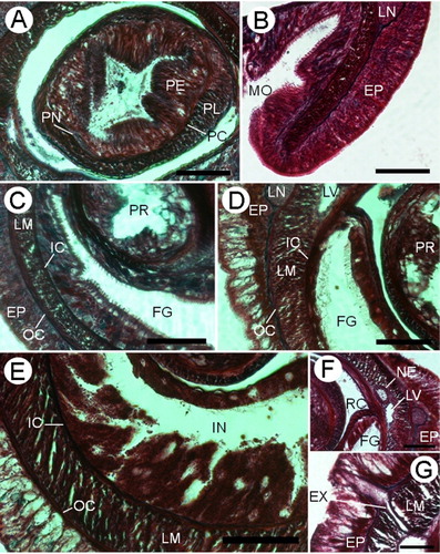
Figure 4 Tubulanus mawsoni (Wheeler, Citation1940) comb. nov.A, Transverse section through the proboscis. B, Transverse section through the mouth showing the buccal chamber. C, Transverse section through the anterior foregut region. D, Transverse section through the posterior foregut region. E, Transverse section through the intestinal region. F, Transverse section through the foregut region showing the arrangement of the nephridial ducts and the lateral blood vessel. G, Transverse section through the foregut region, showing an excretory collecting tubule (EX). EP, epidermis; FG, foregut; IN, intestine; LM, body-wall longitudinal muscle layer; LN, lateral nerve cord; LV, lateral blood vessel; MO, mouth; NE, nephridial duct; OC, body-wall outer circular muscle layer; PE, proboscis outer glandular epithelium; PC, proboscis outer circular muscle layer; PL, proboscis inner longitudinal muscle layer; PR, proboscis; RC, rhynchocoel. Scale bars: 100 µm.
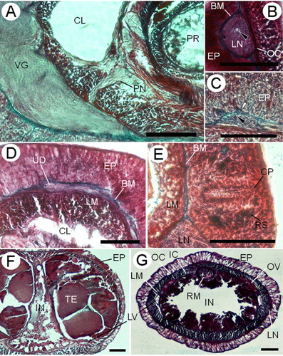
Figure 5 Tubulanus mawsoni (Wheeler, Citation1940) comb. nov.A, Transverse section through the posterior portion of the brain showing the connection between the ventral cerebral commissure and the proboscis nerve. B, Transverse section through the lateral nerve cord showing its position between the outer circular muscle layer and the basal membrane; black arrowhead points to a neurochord. C, Transverse section through the upper mid-dorsal nerve; white arrowhead points to the basal membrane; black arrowhead points to the inner neurilemma. D, Transverse section through the posterior portion of the dorsal cerebral commissure showing the junction with the upper mid-dorsal nerve. E, Transverse section through the cerebral sensory organ pit (CP). F, Transverse section through the intestinal region in the mature male. G, Transverse section through the intestinal region in the immature female. BM, basal membrane; CL, cephalic blood lacuna; EP, epidermis; IC, body-wall inner longitudinal muscle layer; IN, intestine; LN, lateral nerve cord; LM, body-wall longitudinal muscle layer; LV, lateral blood vessel; PN, proboscis nerve; PR, proboscis; OC, body wall outer longitudinal muscle layer; OV, ovum; RM, retractor muscle; RS, deep red-staining cell; TE, testis; UD, upper mid-dorsal nerve; VG, ventral cerebral ganglion. Scale bars: 100 µm.

Character matrix for Tubulanus mawsoni (Wheeler, Citation1940) comb. nov. Character states and code from Sundberg et al. (Citation2009).

Reprints and Corporate Permissions

Please note: Selecting permissions does not provide access to the full text of the article, please see our help page How do I view content?

To request a reprint or corporate permissions for this article, please click on the relevant link below:

Academic Permissions

Please note: Selecting permissions does not provide access to the full text of the article, please see our help page How do I view content?

Obtain permissions instantly via Rightslink by clicking on the button below:

If you are unable to obtain permissions via Rightslink, please complete and submit this Permissions form. For more information, please visit our Permissions help page.