Figures & data
Figure 1. Cranium and premaxilla of kākāpō (Strigops habroptilus; LB14836). A, Left lateral view with rhinotheca in place; B, ventral view with rhinotheca removed to show exposed premaxilla. Photograph: J Froggatt.

Figure 2. Part of rhinotheca of kākāpō (LB14794); the section that lies under the premaxilla (only) with dorsal section removed. ‘L’ is the ledge and ‘S' is the palatal stop against which the gnathotheca works during chewing, ‘a’ points to the vertical surface of the abutment. A, External buccal surface (i.e. ventral view); B, view of hidden internal surface that is normally in contact with the premaxilla; C, proximal end showing heavily-pitted inter-ridge areas; D, left lateral view (bill tip to left); E, view of cross-section of rhinotheca along the mid-line (bill tip to left) showing relative thickness of rhinotheca along its length. Photograph: P Quin.
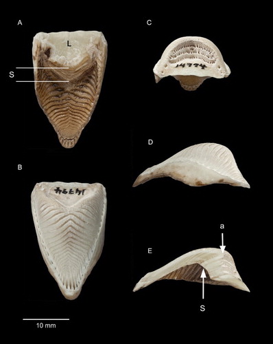
Figure 3. Rhinothecas of kākāpō (A, LB14838), kākā (Nestor meridionalis; B, LB14862) and kea (N. notabilis; C, LB14865). Left lateral view. Photograph: P Quin.
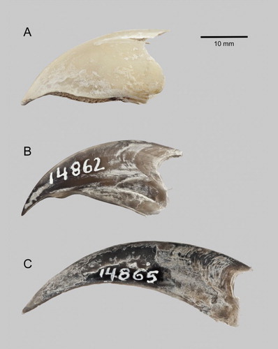
Figure 4. Rhinothecas of kākāpō (A, LB14838), kākā (Nestor meridionalis; B, LB14862) and kea (N. notabilis; C, LB14865). External buccal surface (i.e. ventral view). ‘S1’ is the palatal stop equivalent to that of kākāpō, ‘S2’ is the additional palatal stop of kākā. Photograph: P Quin.
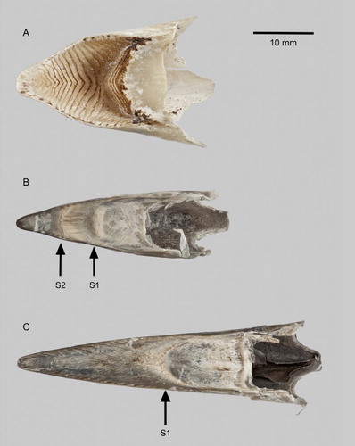
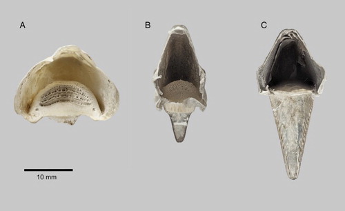
Figure 5. Rhinothecas of kākāpō (A, LB14838), kākā (Nestor meridionalis; B, LB14862) and kea (N. notabilis; C, LB14865). View of proximal end showing cavity after removal of premaxilla. Photograph: P Quin.