Figures & data
Figure 1. 1a: Subcutaneous, diffuse accumulation of fibrinous exudate in and around the keel bursa of a broiler chicken carcass infected with M. synoviae. 1b: Extensive localized accumulation of caseous exudate in the keel bursa of a 47-day-old broiler chicken infected with M. synoviae.
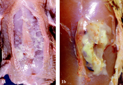
Figure 2. Enlarged liver with greenish tinge and scattered pale reticular pattern from a 47-day-old broiler chicken infected with M. synoviae.
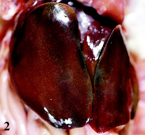
Figure 3. Enlarged mottled spleen of a 47-day-old broiler chicken infected with M. synoviae.
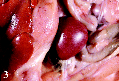
Table 1. Frequency and severity of gross lesions associated with M. synoviae in selected organs of broiler chickens
Table 2. Relative frequency and severity of microscopic lesions associated with M. synoviae in selected organs of broiler chickens
Figure 4. 4a: Keel bursa from a 47-day-old broiler chicken infected with M. synoviae showing severe perivascular cuffing (arrows) by mononuclear inflammatory cells. Haematoxylin and eosin, bar=200 μm. 4b:Higher magnification of one field from 4a showing in more detail prominent endothelial cells and infiltration by lymphocytes, a few plasma cells and macrophages in and around a blood vessel. Haematoxylin and eosin, bar=50 μm.
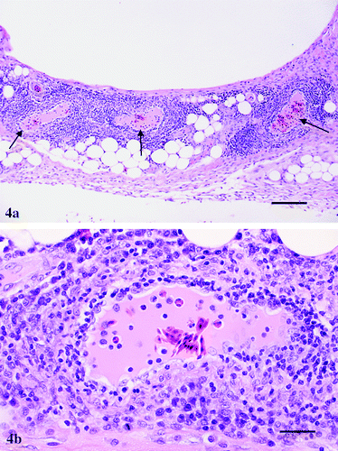
Figure 5. Liver from a 47-day-old broiler chicken infected with M. synoviae, showing prominent and vacuolated endothelial cells and infiltration by lymphocytes, a few plasma cells and macrophages in and around a blood vessel. Haematoxylin and eosin, bar=75 μm.
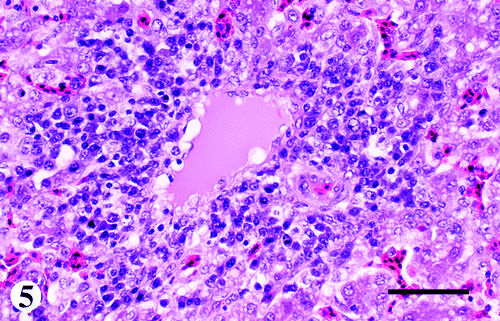
Figure 6. Brain from a 47-day-old broiler chicken infected with M. synoviae, in which there is infiltration of primarily lymphocytes in and around the blood vessel. Haematoxylin and eosin, bar=85 μm.
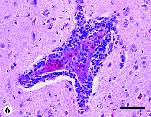
Figure 7. Choroid (*) of the eye from a 47-day-old broiler chicken infected with M. synoviae, in which there is infiltration of lymphocytes mixed with a few plasma cells. Haematoxylin and eosin, bar=100 μm.
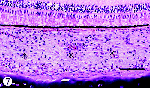
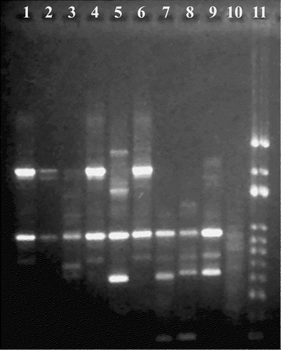
Figure 8. RAPD analysis of M. synoviae isolates. Lanes 1 and 2, isolates from 47-day-old broiler chickens; lanes 3 and 4, isolates from 33-day-old to 39-day-old broiler chickens; lane 5, isolate from a turkey; lane 6, isolate from a broiler breeder; lane 7, MS-H reference strain; lane 8, WVU 1853 reference strain; lane 9, F10-2AS reference strain; lane 10, negative control; lane 11, 100 base pair ladder.