Figures & data
Figure 1. Number of FFU of MDV isolated in CKC, CEKC, and CEF cultures inoculated with 0.5×10⁶ splenocytes/culture infected with RB-1B (1a) or RK-1 (1b). The FFU represent the average for four cultures/bird. Foci were enumerated at 4 d.p.i. (open bars) and 6 d.p.i. (closed bars). The absence of a bar indicates that foci were not present.
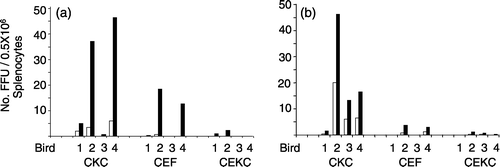
Figure 2. Number of FFU isolated in individual CKC cultures inoculated with 0.5×10⁶ RB-1B-infected or RK-1-infected splenocytes/culture using the results from experiments 2 and 3 (birds 2 to 4 from and , respectively). Each symbol represents one culture/bird. Foci were enumerated at 6 d.p.i.
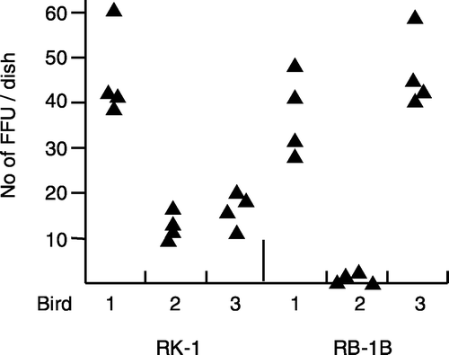
Figure 3. Number of FFU isolated in CKC, CEKC, and CEF cultures inoculated with 0.5×10⁶ cells/culture of tumour material (p2 in CKC) from a Silkie hen (sample 1) and NC tumour cells (sample 2). The FFU represent the average for four cultures/sample. Foci were enumerated at 4 d.p.i. (open bars) and 6 d.p.i. (closed bars). The absence of a bar indicates the absence of foci.
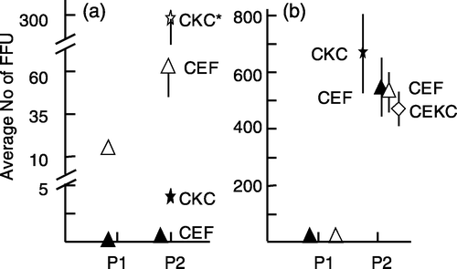
Figure 4. Relative increase of the number of MDV FFU in p2. 4a: p1 RB-1B-infected CKC were trypsinized and the cells were passed to CKC or CEKC (experiment 2). 4b: p1 RK-1-infected CKC were trypsinized and the cells were passed to CKC, CEF, or CEKC (experiment 3). The values are based on the average counts of FFU for three individual bird samples and are expressed as the relative number of FFU in p2 compared with the number of FFU in p1. The average number of FFU for the three birds in p1 was set at 1. The open and closed symbols in (b) are representing the same birds in p1, but cells were passed to CKC and CEKC (open symbols) or CKC and CEF (closed symbols). Note that the scales for (a) and (b) are different. Significant differences: * P<0.05 and *** P<0.001 using Student's t test; vertical bars indicate the standard deviation.
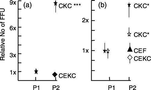
Figure 5. Increase in the number of MDV FFU in p2 using MDV-infected CEKC. 5a: p1 RB-1B-infected CEKC were trypsinized and the cells were passed to CEKC or CKC (experiment 2). 5b: p1 RK-1-infected CEKC were trypsinized and the cells were passed to CEKC, CEF, or CKC (experiment 3). The values represent the average number of FFU based on four dishes/bird for two individual bird samples (#1=open symbols and #2=closed symbols (a) or the average value for three individual birds (b). The open and closed symbols in (b) are representing the same birds in p1, but cells were passed to CEKC and CEF (open symbols) or CEKC and CKC (closed symbols). Significant differences: ** P<0.01 using Student's t test; vertical bars indicate the standard deviation.
Figure 6. Increase in the number of MDV FFU in p2 using MDV-infected CEF. 6a: p1 RB-1B-infected CEF were trypsinized and the cells were passed to CEF or CKC (experiment 2). 6b: p1 RK-1-infected CEF were trypsinized and the cells were passed to CKC, CEF, or CEKC (experiment 3). The values represent the average number of FFU based on four dishes for two individual bird samples (#1=open symbols and #2=closed symbols (a) or the average value for three individual birds (b). The open and closed symbols in (b) are representing the same birds in p1 but cells were passed to CEF and CEKC (open symbols) or CEF and CKC (closed symbols). Significant differences: * P<0.05 using Student's t test; vertical bars indicate the standard deviation.