Figures & data
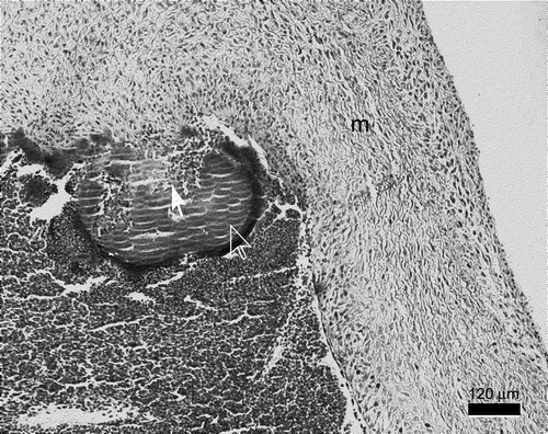
Figure 1. Growth curves of S. gallinaceus (C13156) and E. hirae (C17410) expressed as optical density (OD 620 nm) and corresponding viable counts (log10 CFU/ml). Inocula used for infection were adjusted to an OD 620 nm of 0.1 in PBS (approximately 108 CFU/ml) taken from exponential growth of culture (approximately 9 h).
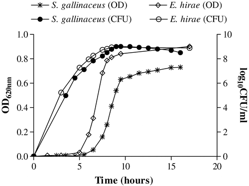
Table 1. Recovery of S. gallinaceus post mortem from birds experimentally infected via the brachial vein
Figure 2. Gross postmortem lesions following S. gallinaceus jugular intravenous infection. 2a: hepatomegaly. 2b: splenomegaly with multiple, randomly distributed, irregular, well-demarcated areas of necrosis surrounded by a haemorrhagic zone (arrow). 2c: yellow–white vegetative valvular endocarditis (arrow).
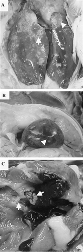
Table 2. Recovery of S. gallinaceus post mortem from birds experimentally infected via the jugular vein
Table 3. Recovery of E. hirae and lesions seen post mortem in birds experimentally infected via the brachial vein
Figure 3. Gross postmortem lesions following E. hirae jugular intravenous infection. 3a: hepatomegaly. 3b: splenomegaly with multiple, randomly distributed, irregular, well-demarcated areas of necrosis surrounded by a haemorrhagic zone (arrow). 3c: yellow–white lesion of vegetative valvular endocarditis (arrow denotes thickening of the mitral valve due to vegetative growth).
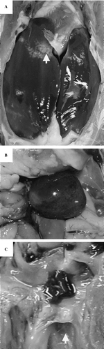
Table 4. Recovery of E. hirae and lesions seen post mortem in birds experimentally infected via the jugular vein
Figure 4. Percent of positive cultures post mortem from specific tissues from birds at times post infection with (4a) S. gallinaceus via the brachial vein, (4b) E. hirae via the brachial vein, (4c) S. gallinaceus via the jugular vein, and (4d) E. hirae via the jugular vein.
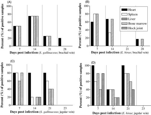
Figure 5. Mitral valve endocarditis induced by S. gallinaceus 3 weeks post-infection. The bird had hepatomegaly and splenomegaly with necrosis, renomegaly and both valvular and mural endocarditis. There is formation of a vegetative lesion at the mitral valve (m) consisting of bacteria, fibrin and basophils (full arrow), surrounded by a corona consisting mainly of heterophils and erythrocytes in the periphery (open arrow).