Figures & data
Table 1. Mycoplasma strains used in M. gallisepticum Q-PCR cross-reactivity testing
Figure 1. Reproducibility of Q-PCR crossing points of DNA standards compared with log10 M. gallisepticum CFU/ml determined by culture. Mean of five different Q-PCR runs. Bars indicate SEM.
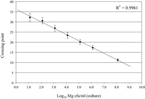
Figure 2. Results of M. gallisepticum DNA standard in Q-PCR and electrophoresis. 1 to 7, 10-fold dilutions from culture A, tested in duplicate, from undiluted (sample 1, 106.1 CFU equivalents/ml) to 10−6 (sample 7, 101.1 CFU equivalents/ml); 8, H2O; M, marker (DNA 50 bp Marker XIII; Roche Applied Science, Mannheim, Germany).
Table 2. Results of reproducibility of Q-PCR based on M. gallisepticum DNA standards derived from culture A
Table 3. Results of Q-PCR and culture on time-interval samples taken from culture B
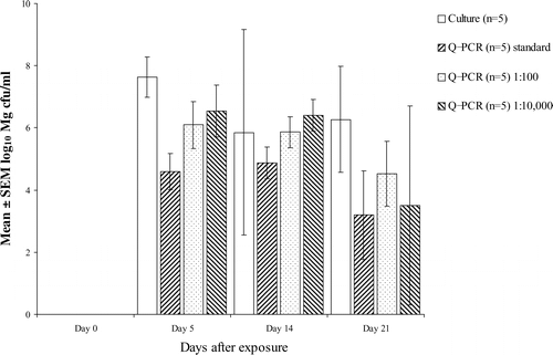
Figure 3. Effect of dilution of samples on estimation of CFU equivalents/ml using Q-PCR (samples from experiment B). Bar indicates SEM.