Figures & data
Figure 1. Large white foci of necrosis (arrow) in the heart from a dead duck. Bar = 0.6 cm.
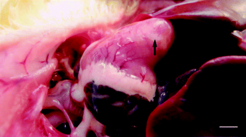
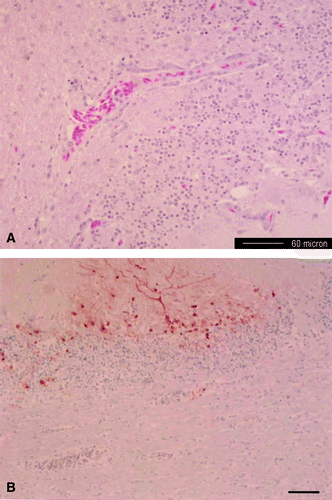
Figure 2. Photomicrograph of the cerebellum from a dead duck. 2A: focal mononuclear perivascular cuffs with gliosis in the neurophil. Haematoxylin & eosin stain. Bar = 60 µm. 2B: viral antigen detected in the Purkinje neurons, dendrites and granular cell layer. Immunohistochemical stain with haematoxylin counterstain. Bar = 75 µm.